Monday, June 22, 2026
microfluidics WIP
PermalinkExperiment Gallery
Daily Notes
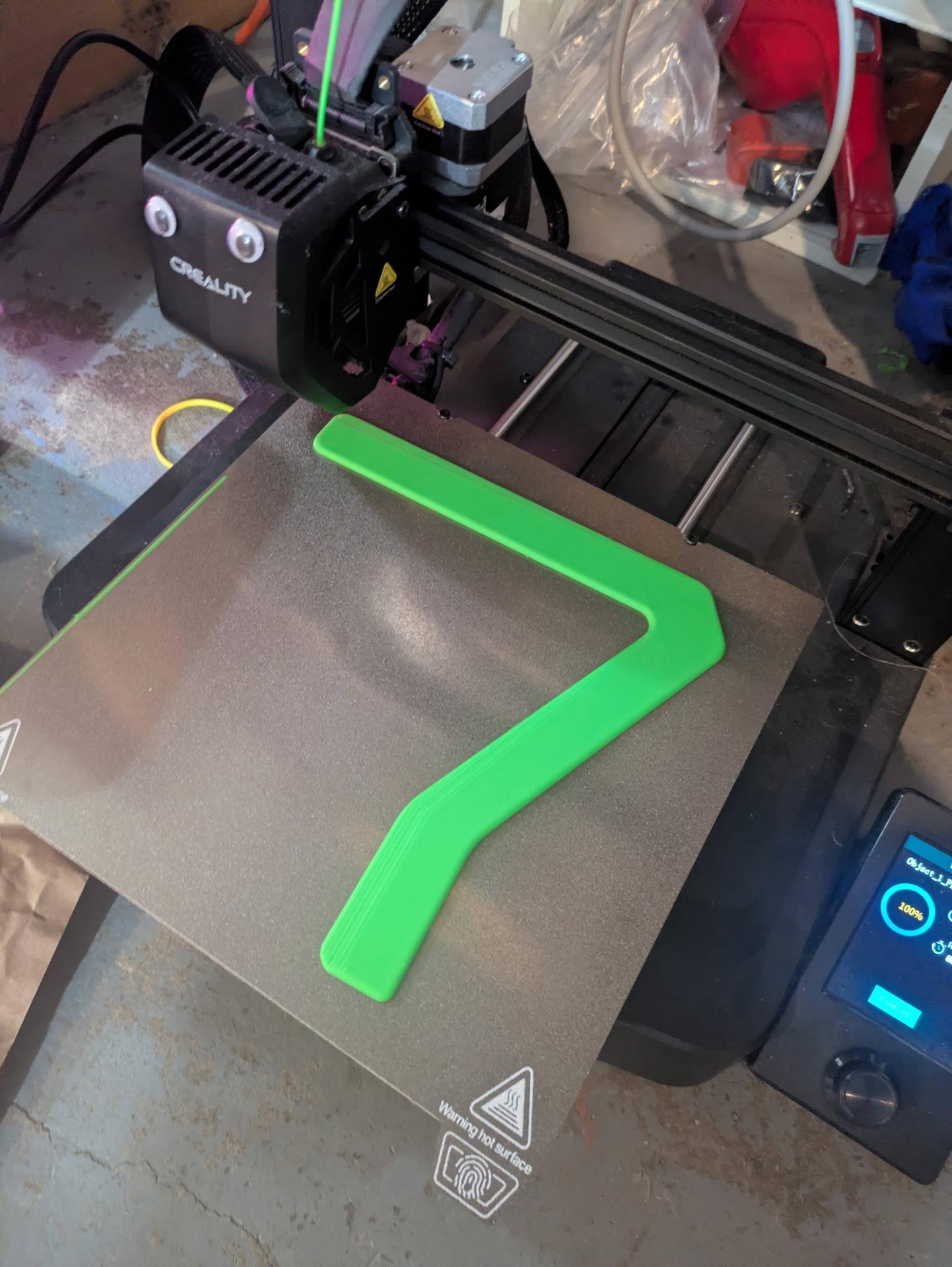
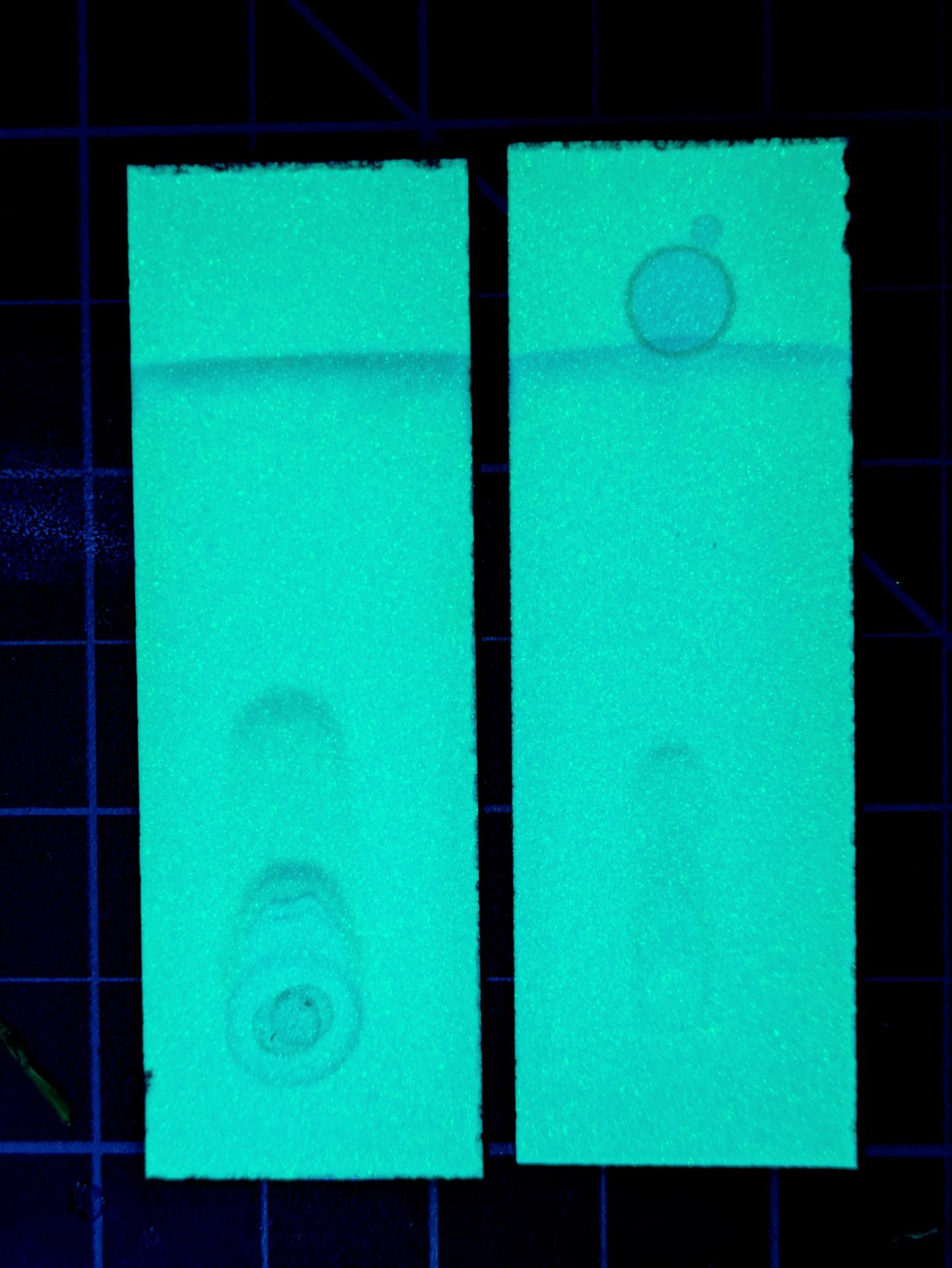
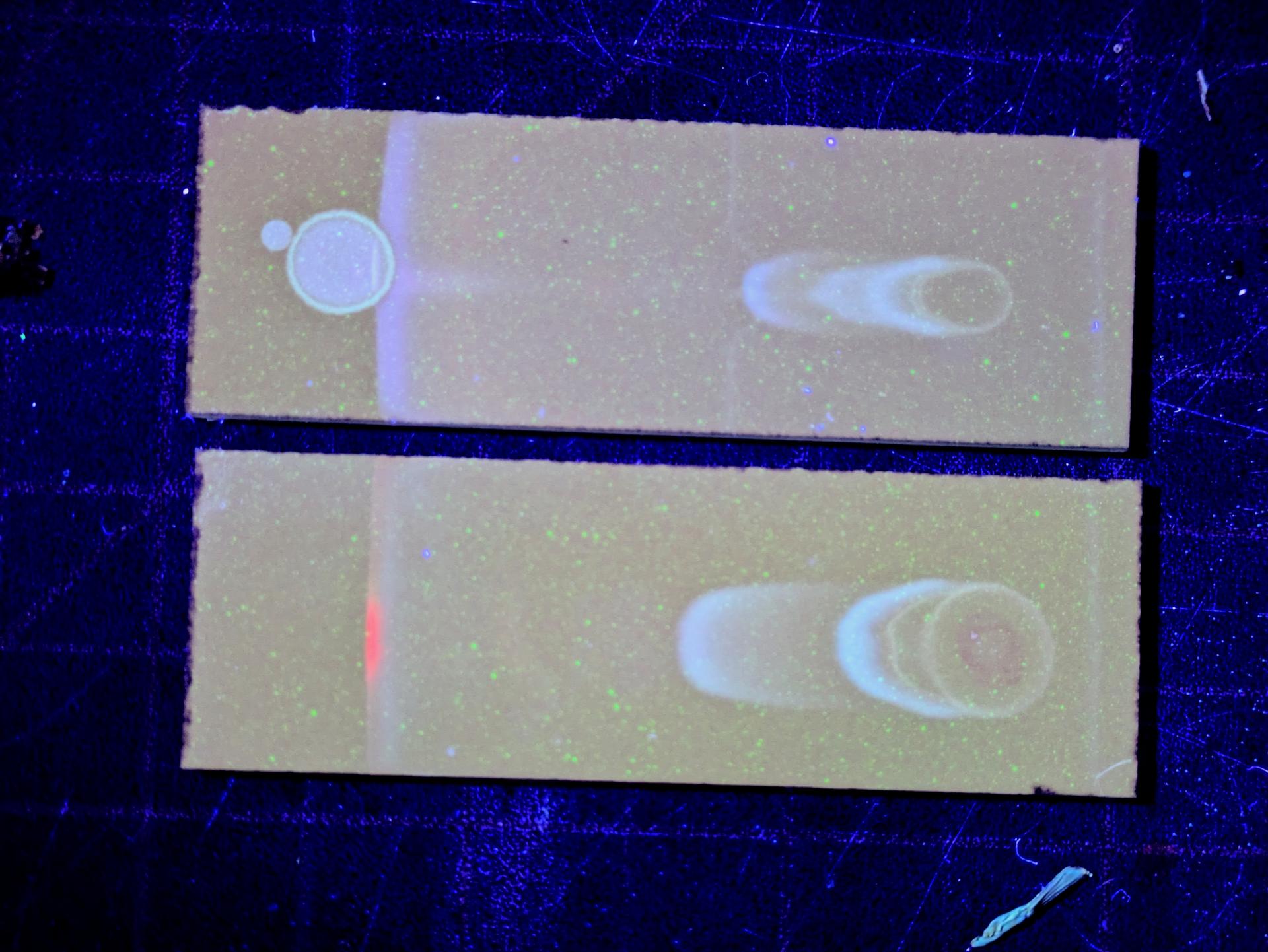
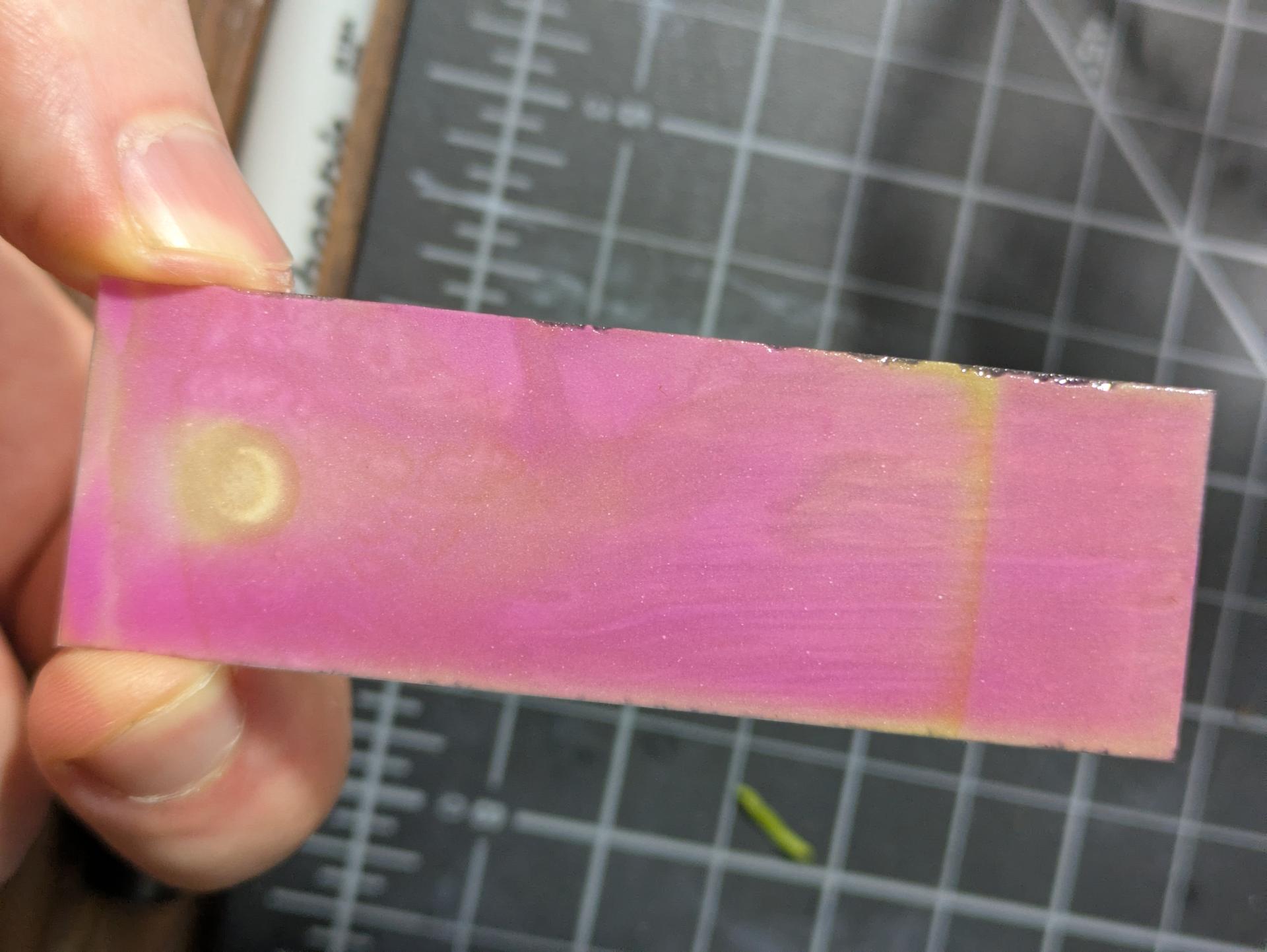
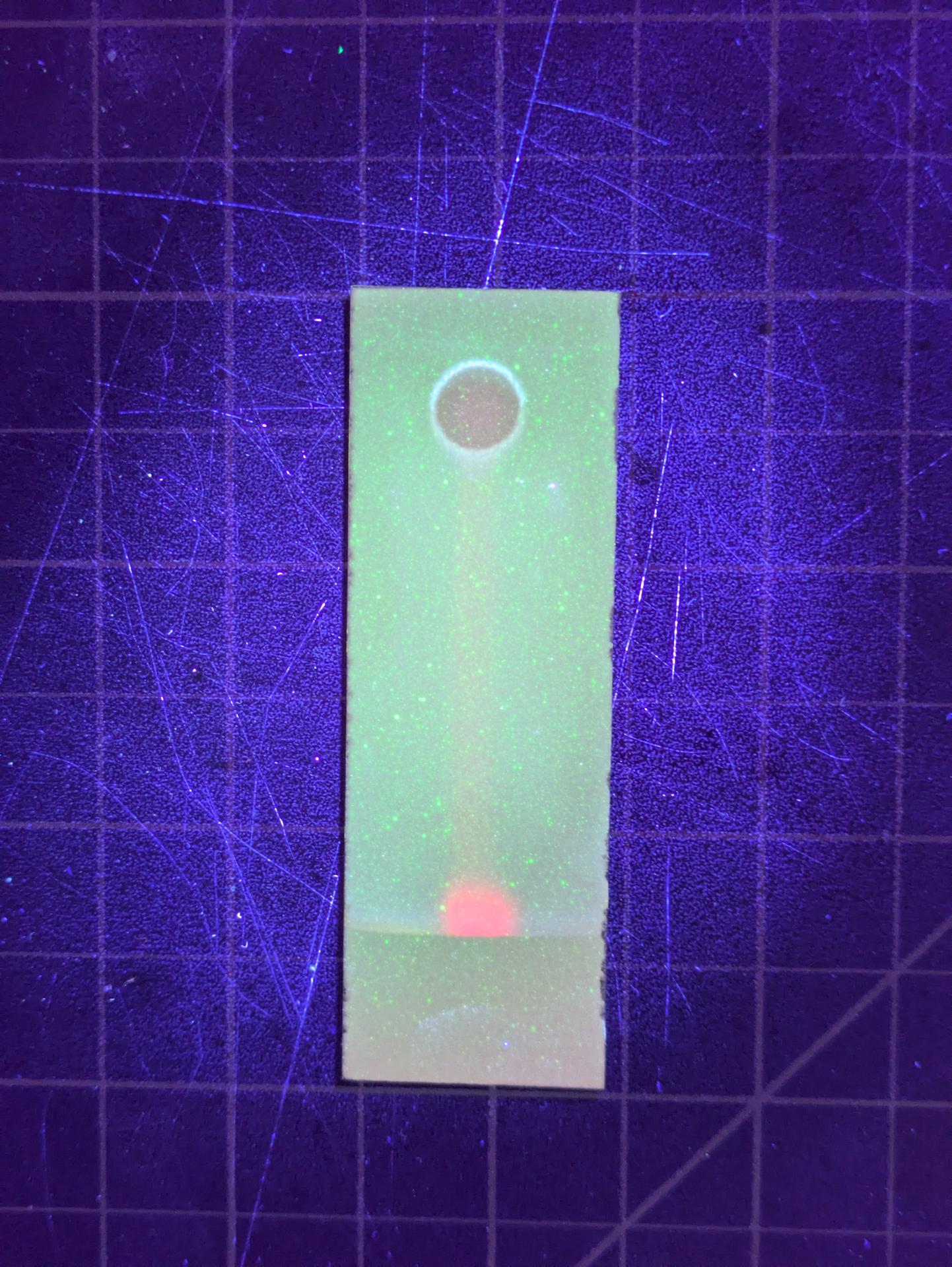
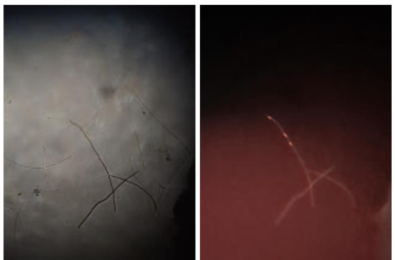
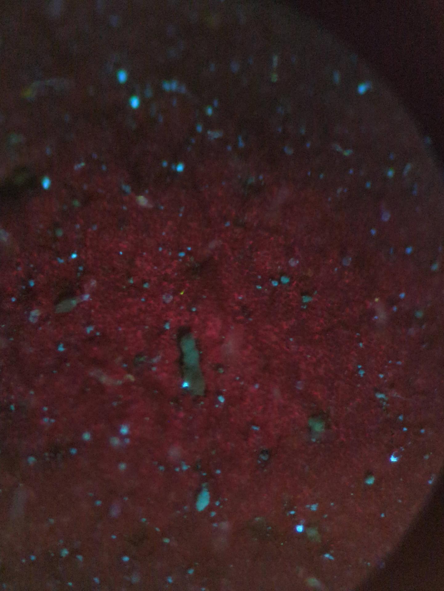
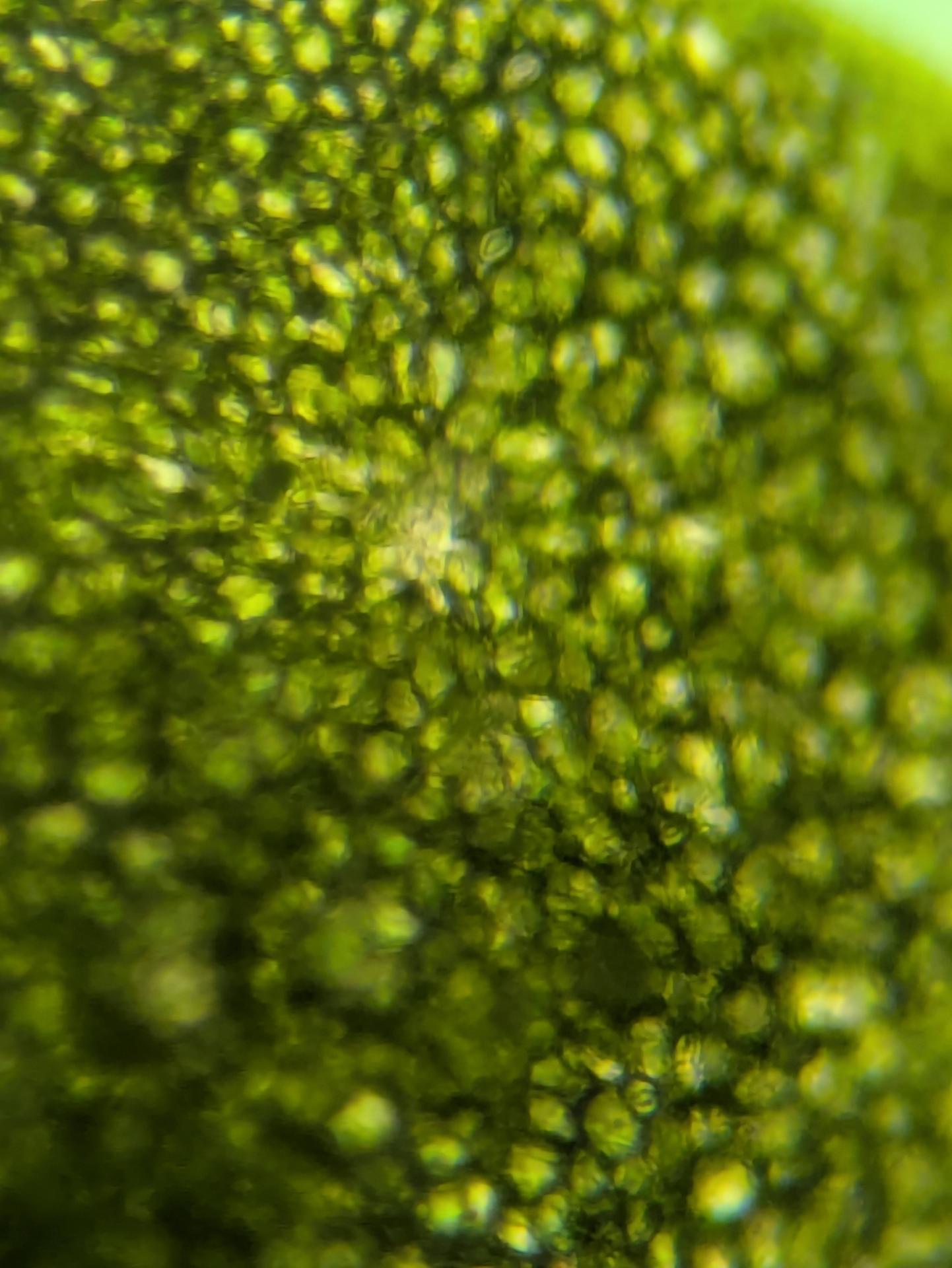
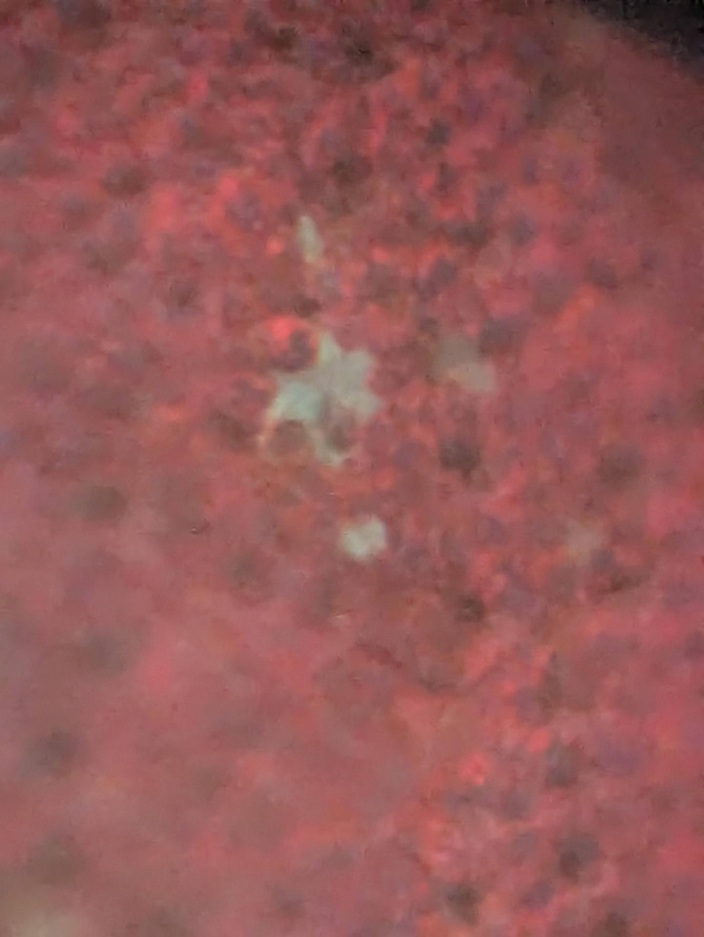
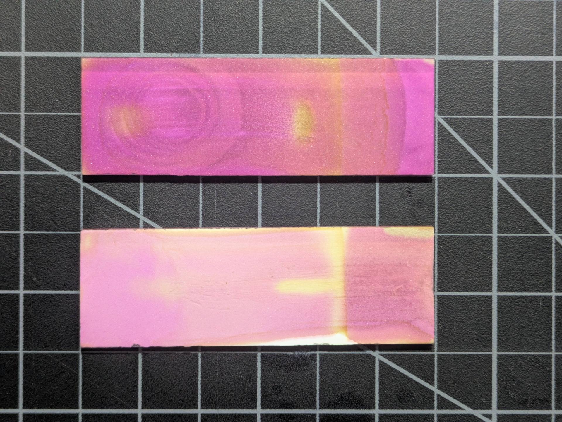
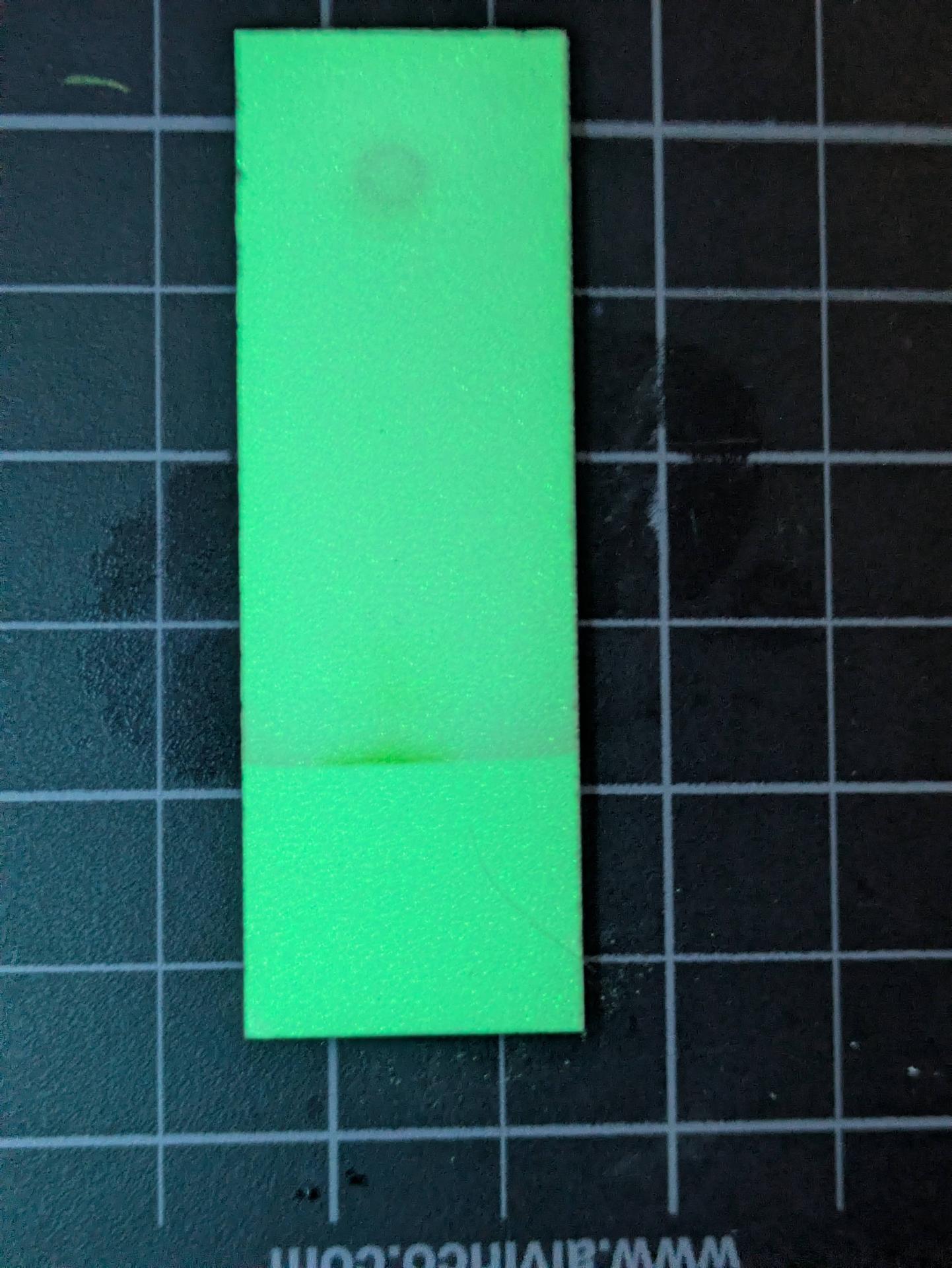
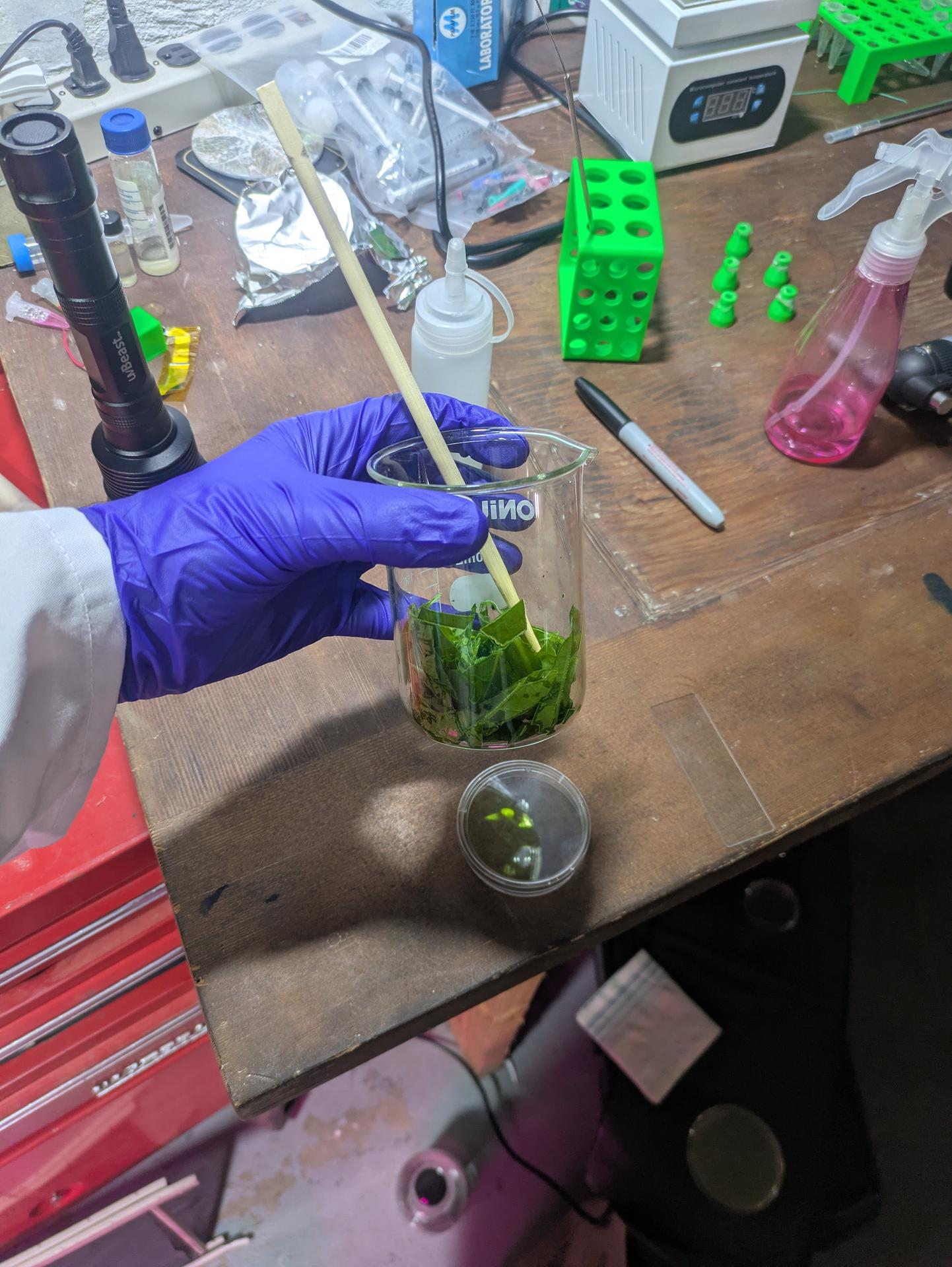
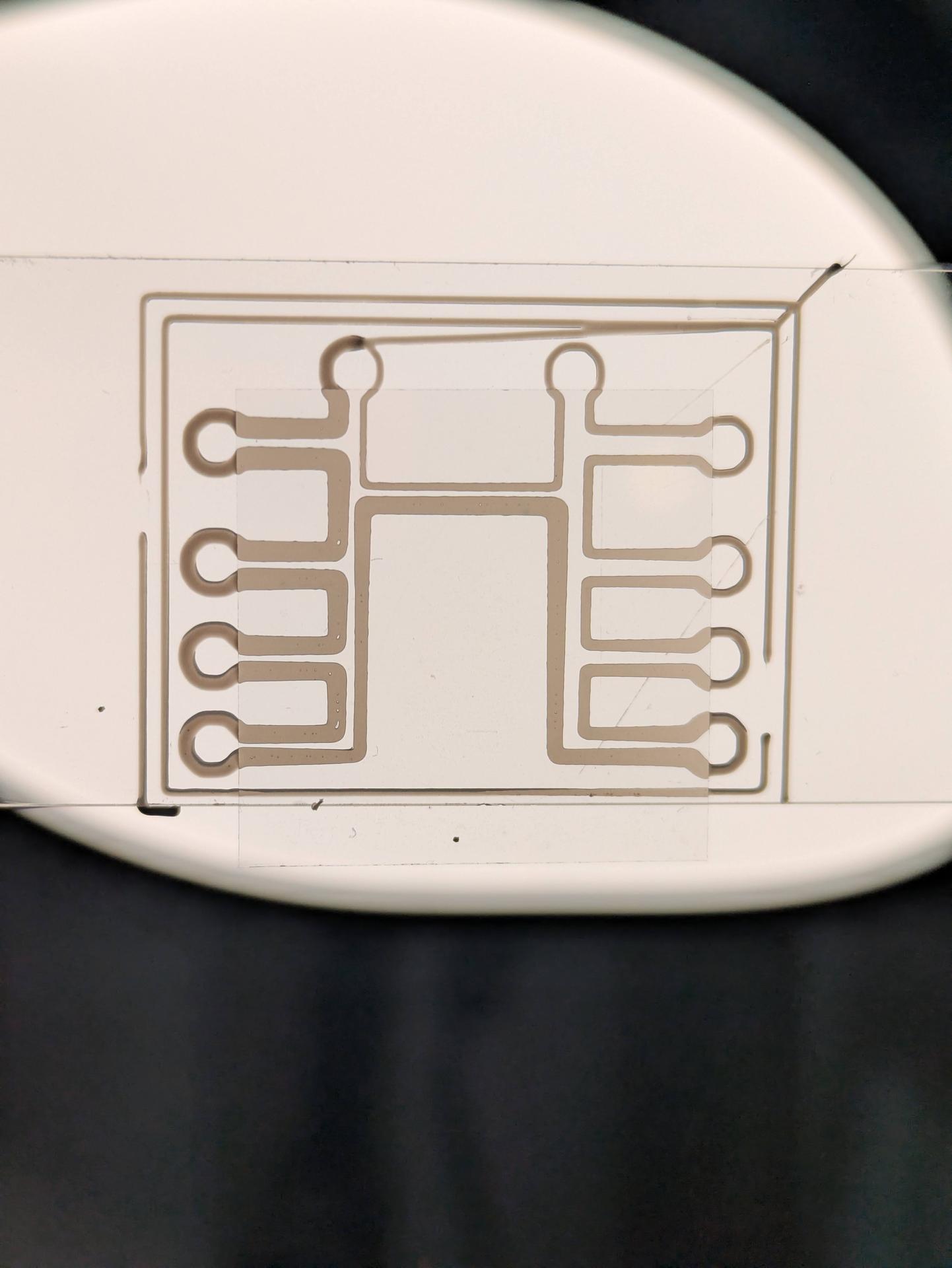
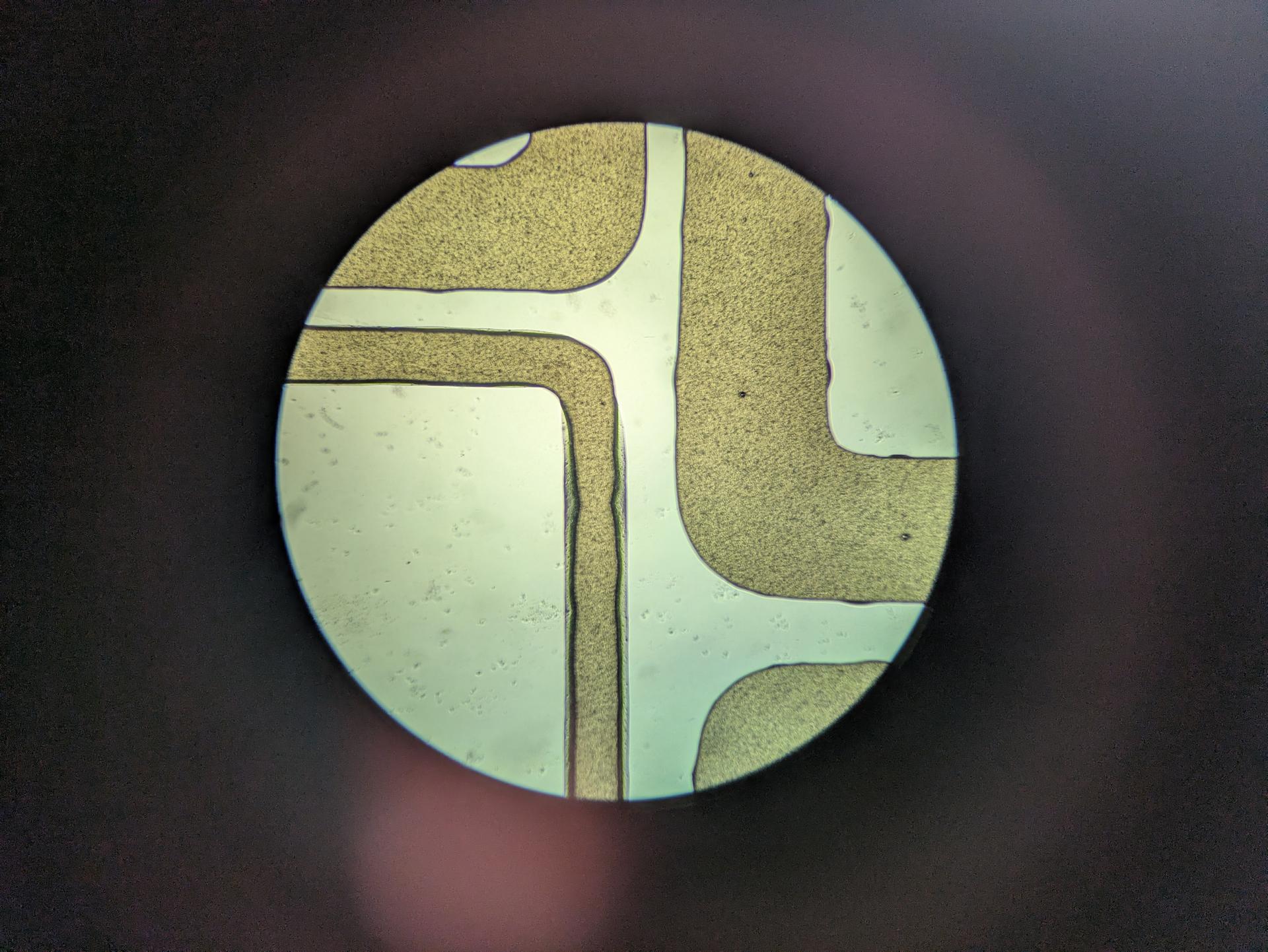
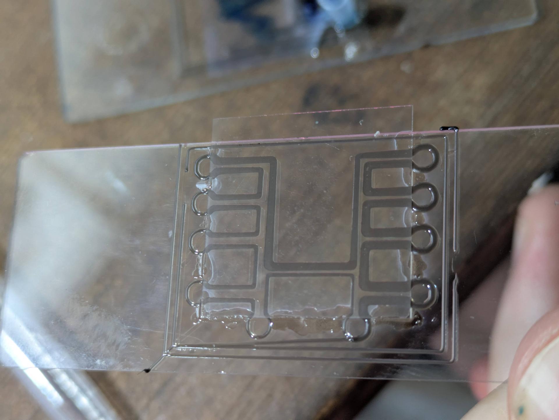
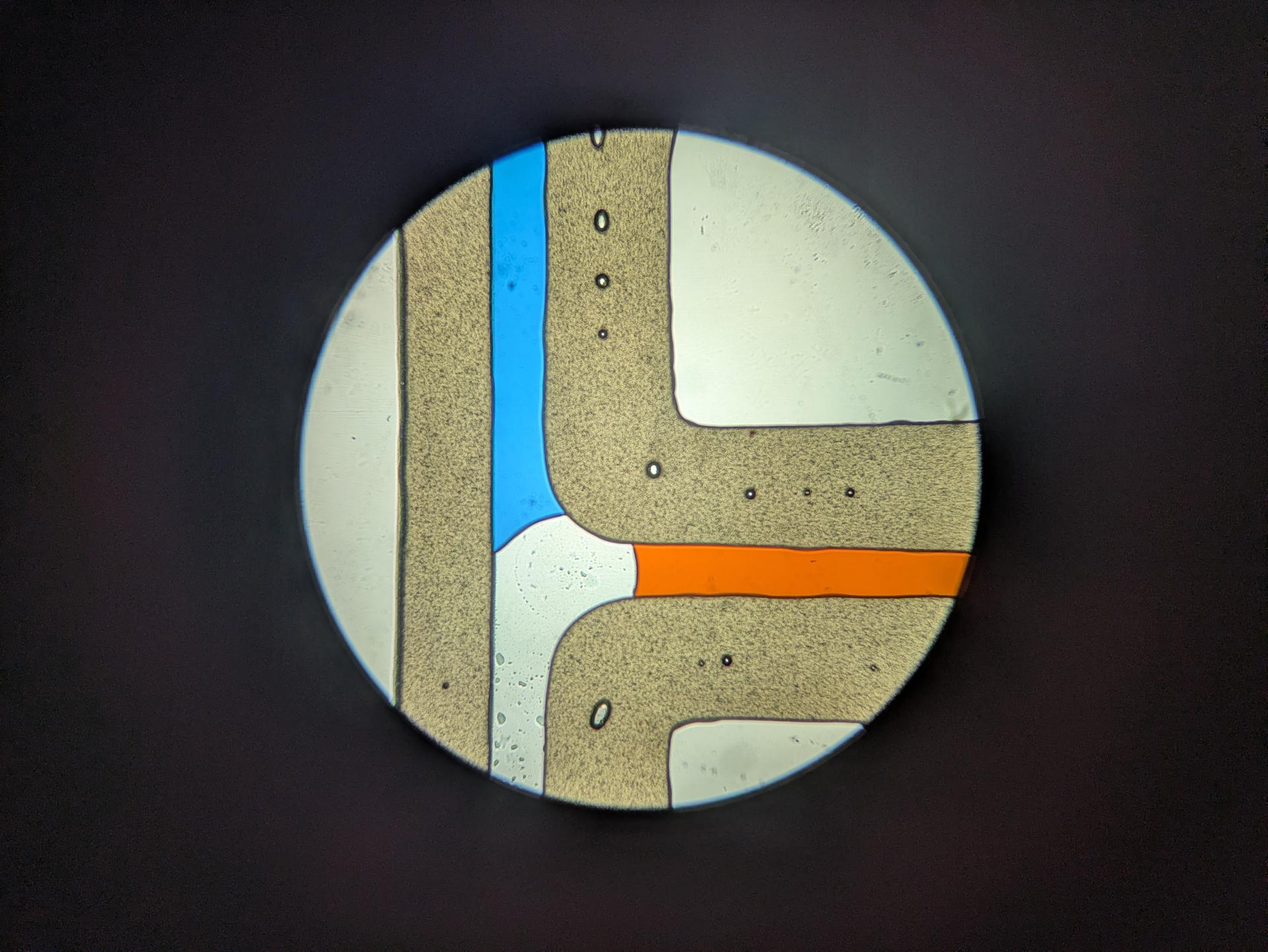
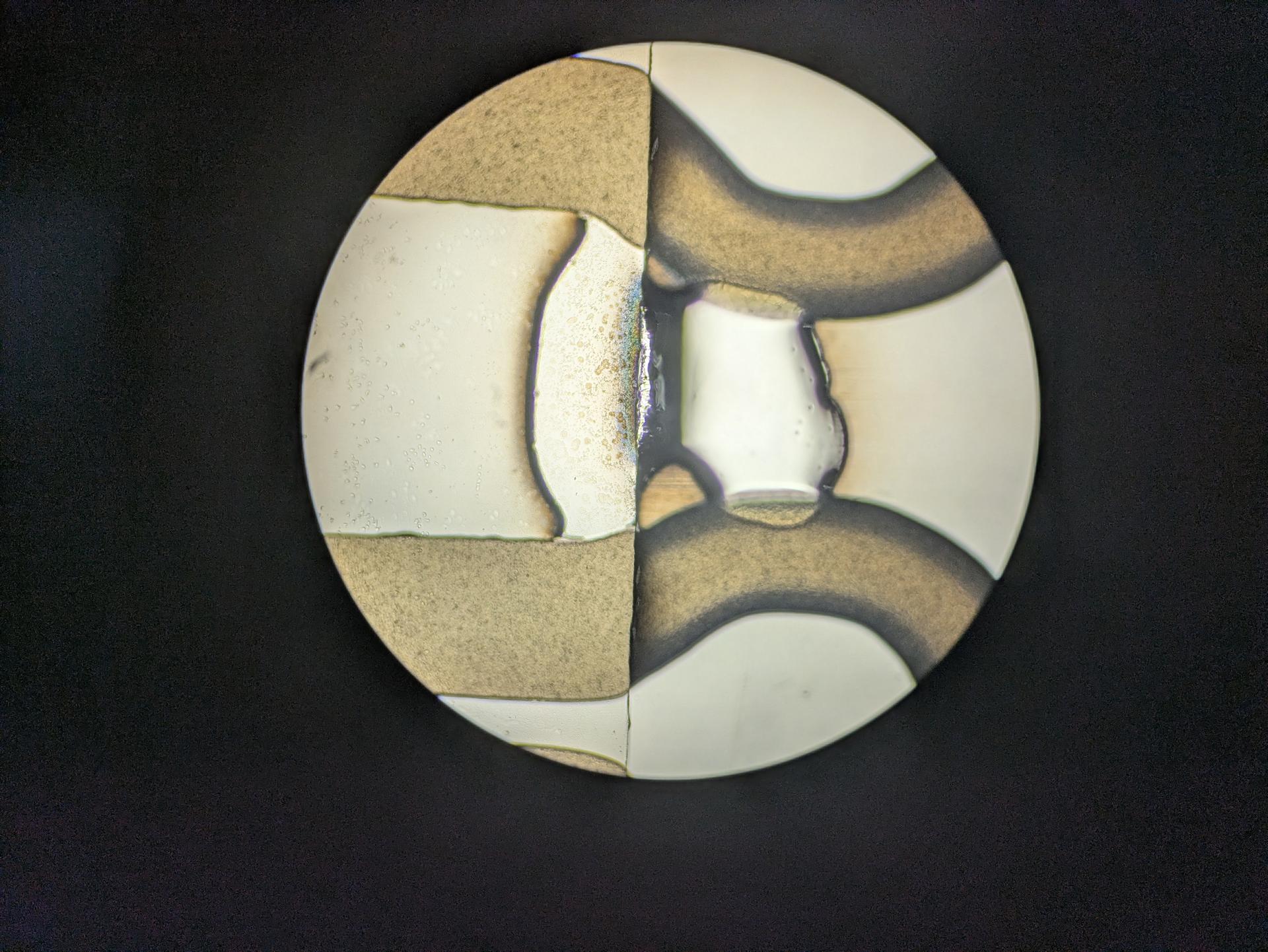
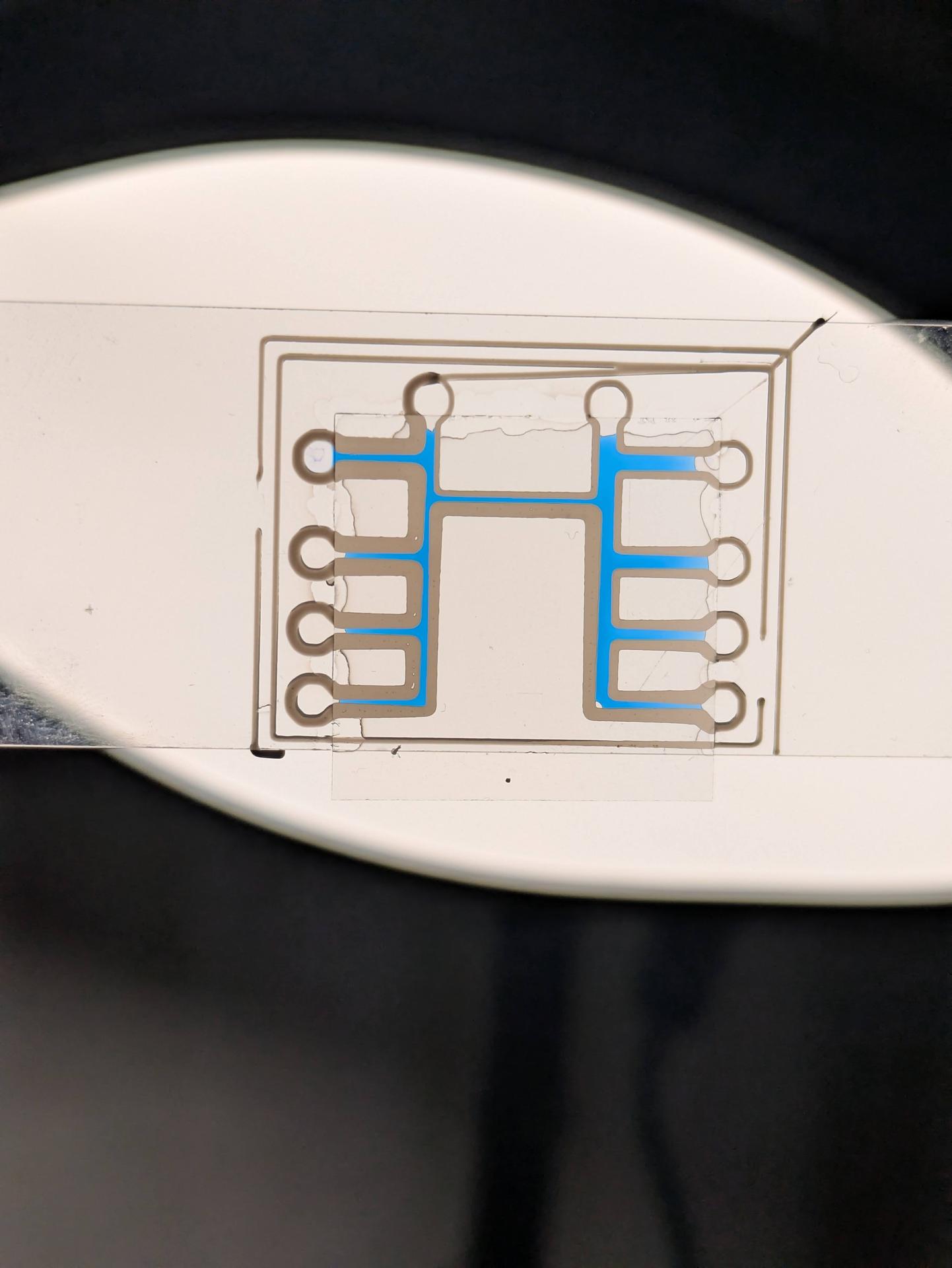
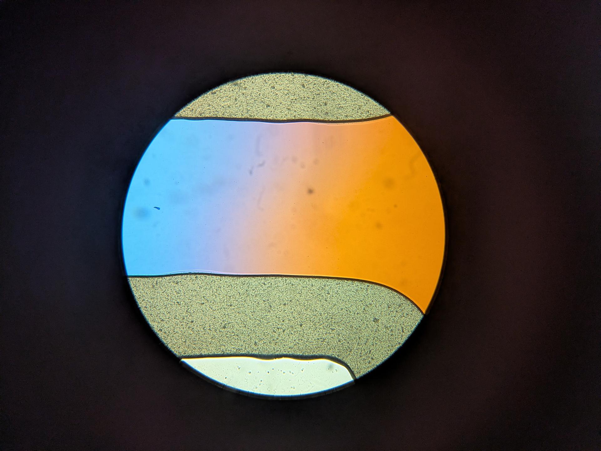
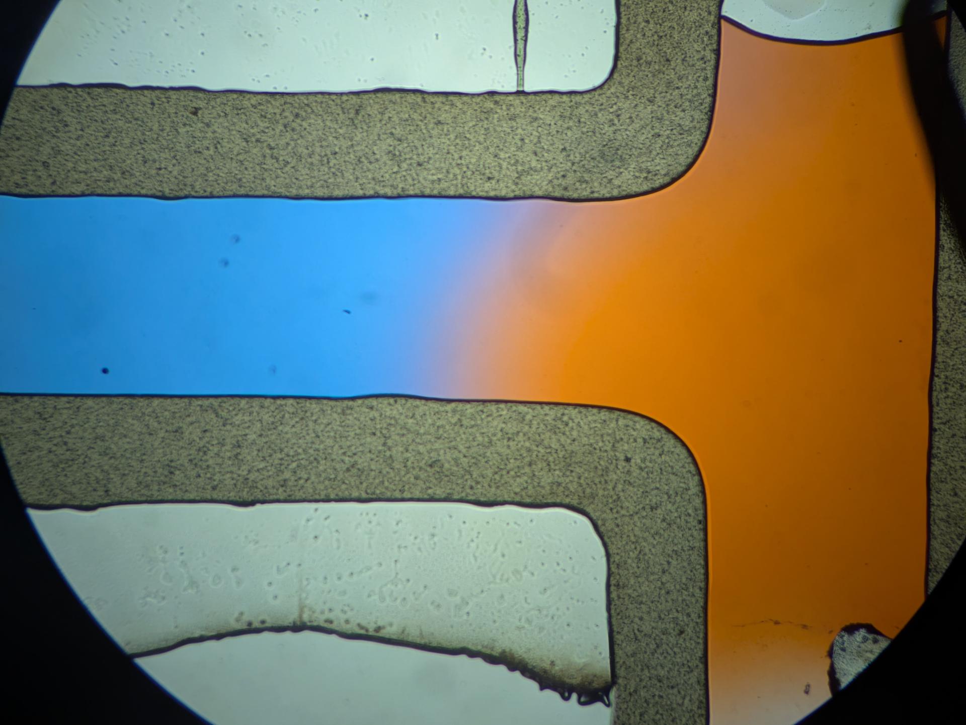
I have just started testing laying down PLA on a glass slide to make microfluidic channels - or maybe just 'fluidic', since the smallest gap I've done is 100um. The main trick is placing a cover slip on top and then setting it on a hot plate set to 180C until the pla melts and forms a sealed surface with the slide+coverslip. Don't leave too long (everything will smush and fuse) but with right timing you can get very nice chips. Make sure glass is clean. I have script I'm tweaking for custom gcode gen. Will obv post more. For now, glamor shots of early tests: