Open Science Journal

Chronological Lab Notebook

Daily logs with notebook pages, experiment photos, and markdown notes.

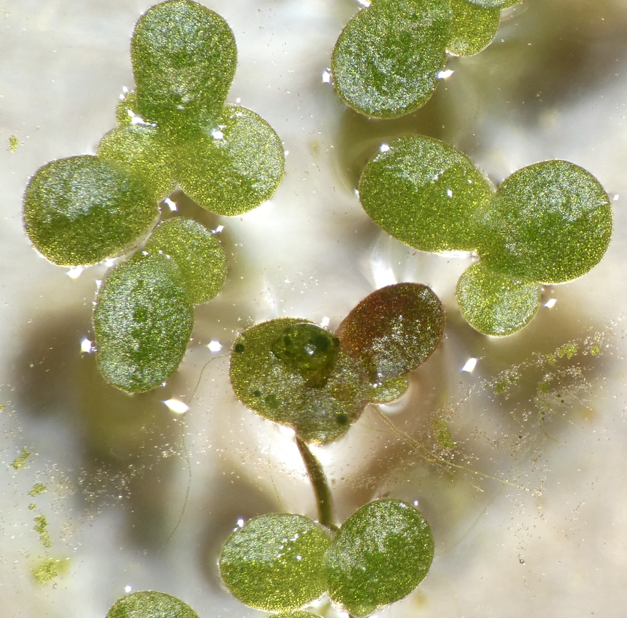
Transformed Duckweed
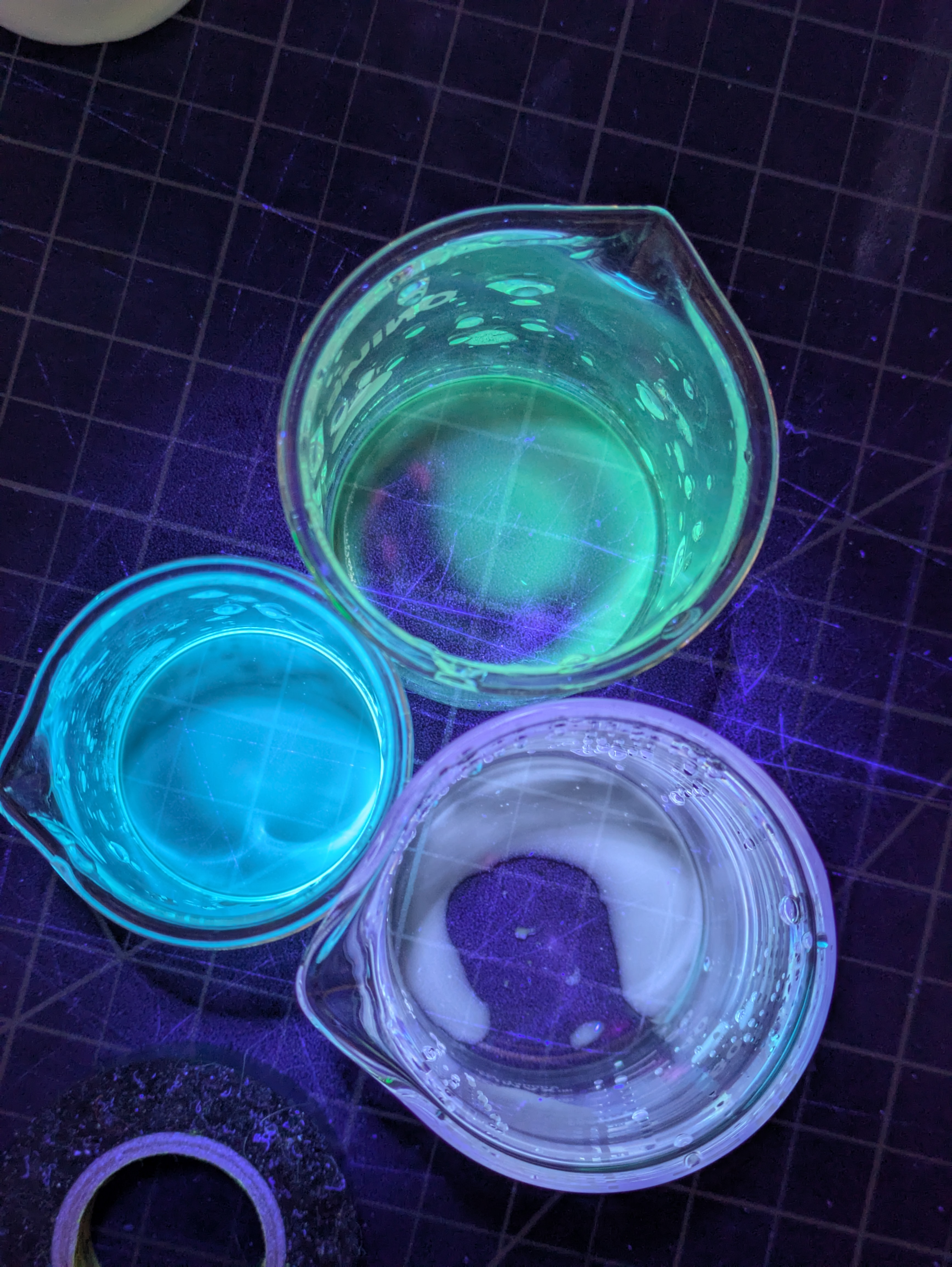
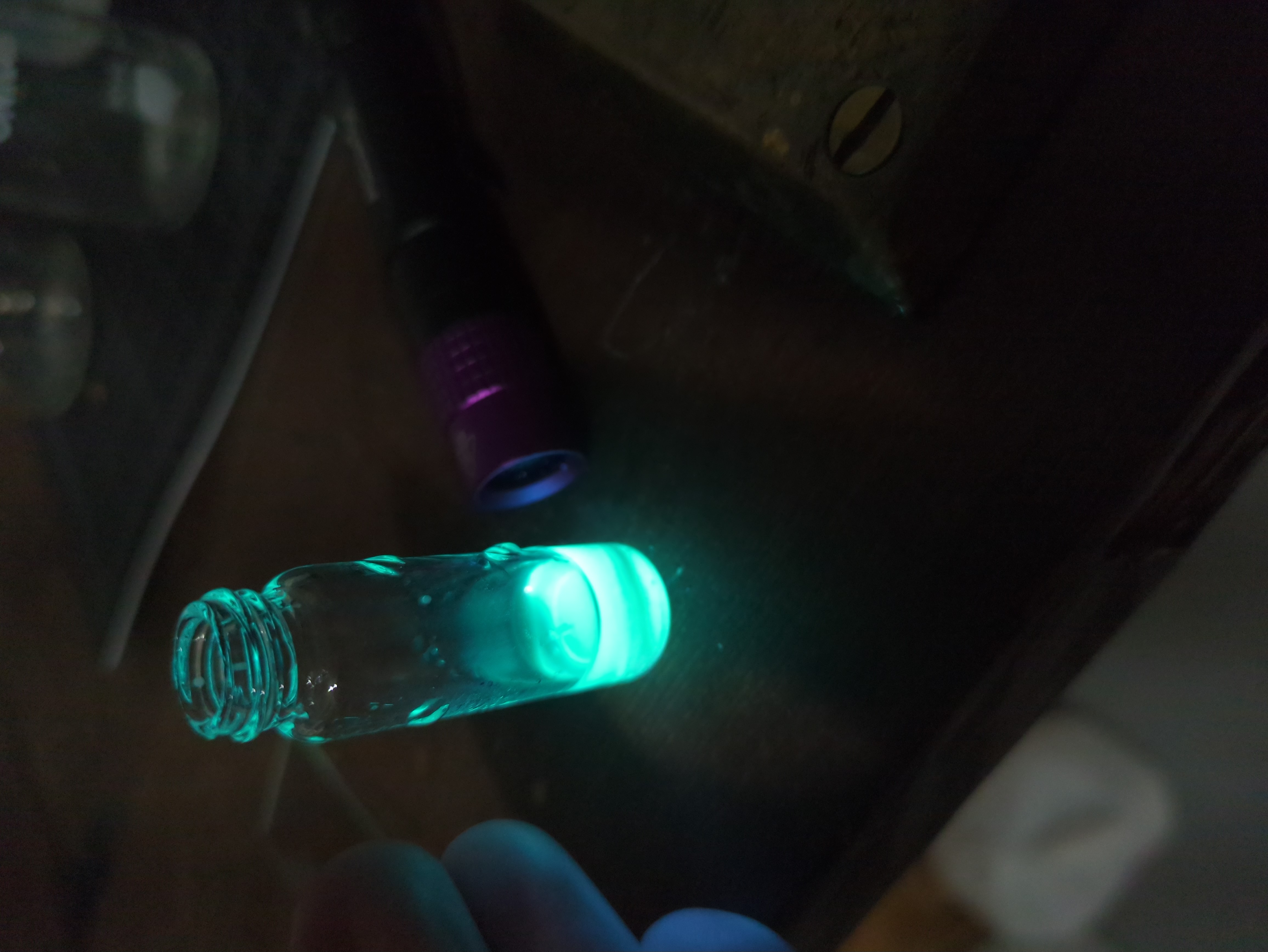
Permalink

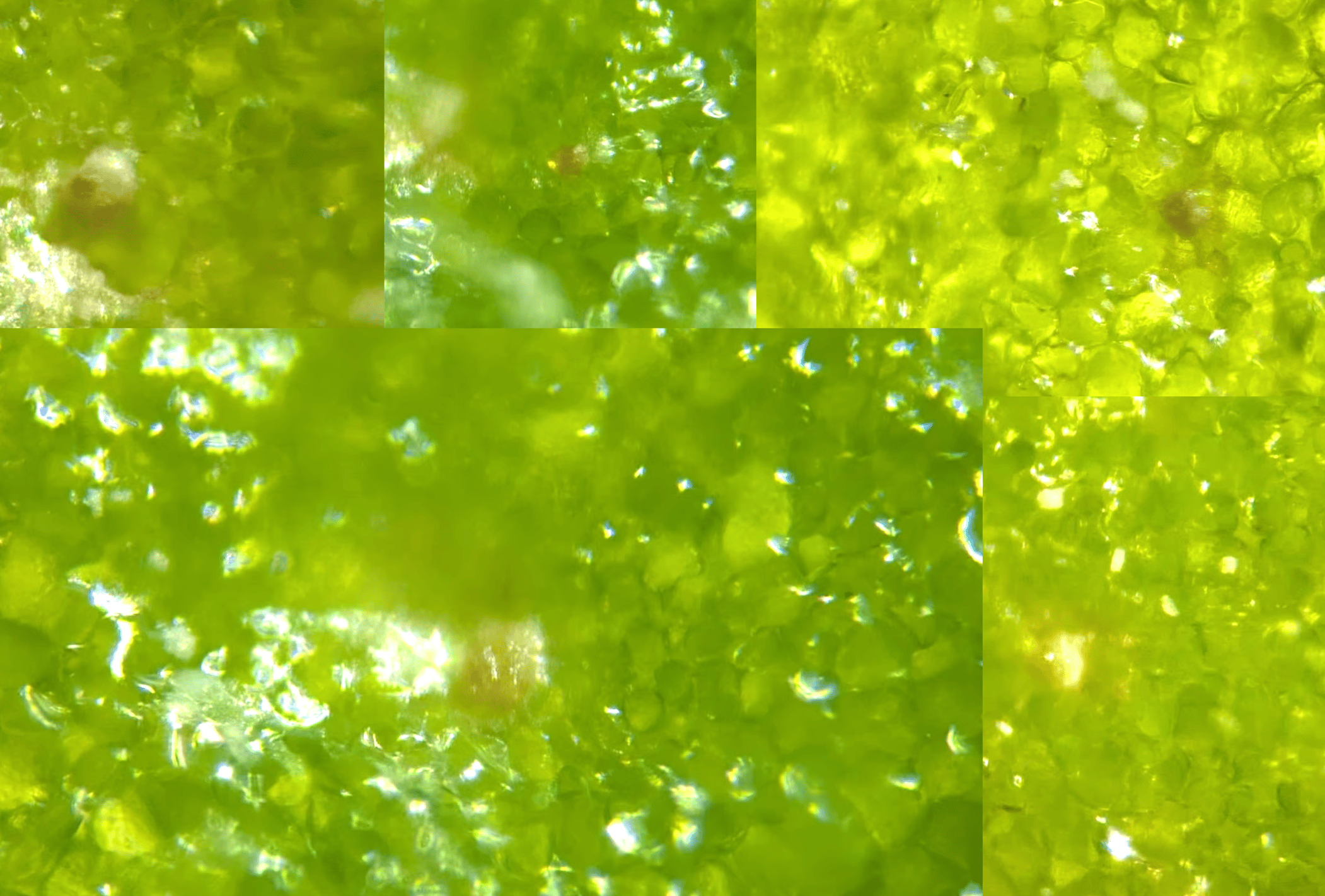
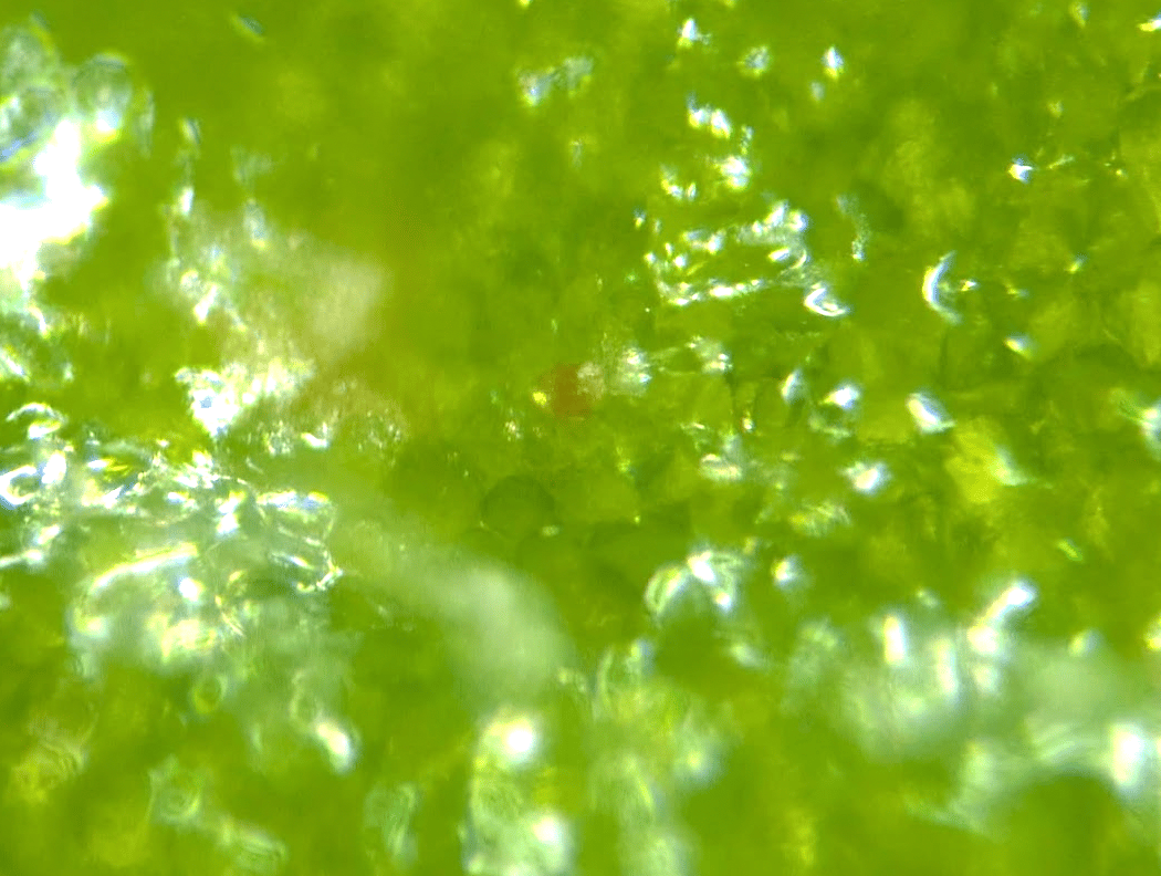
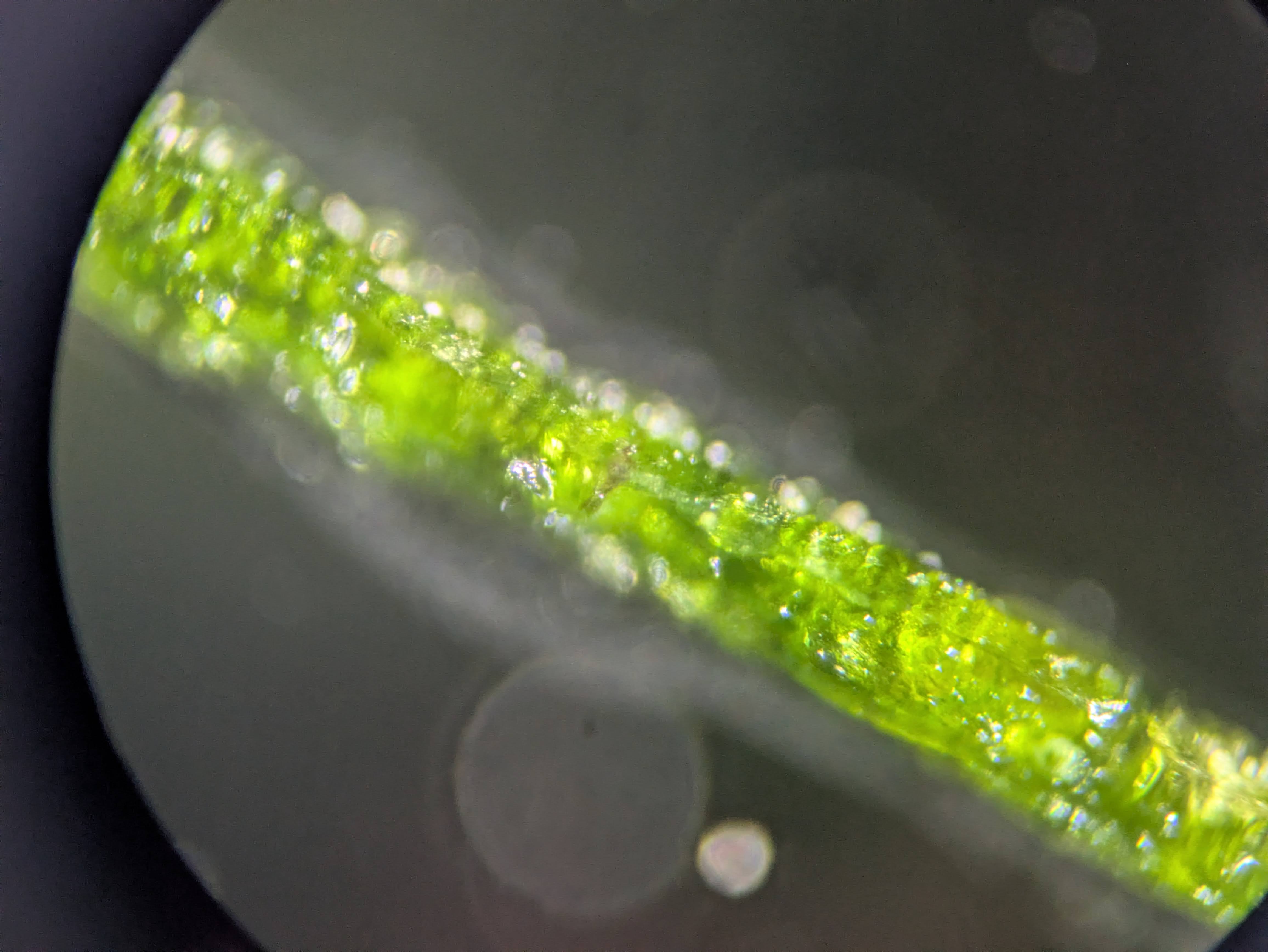
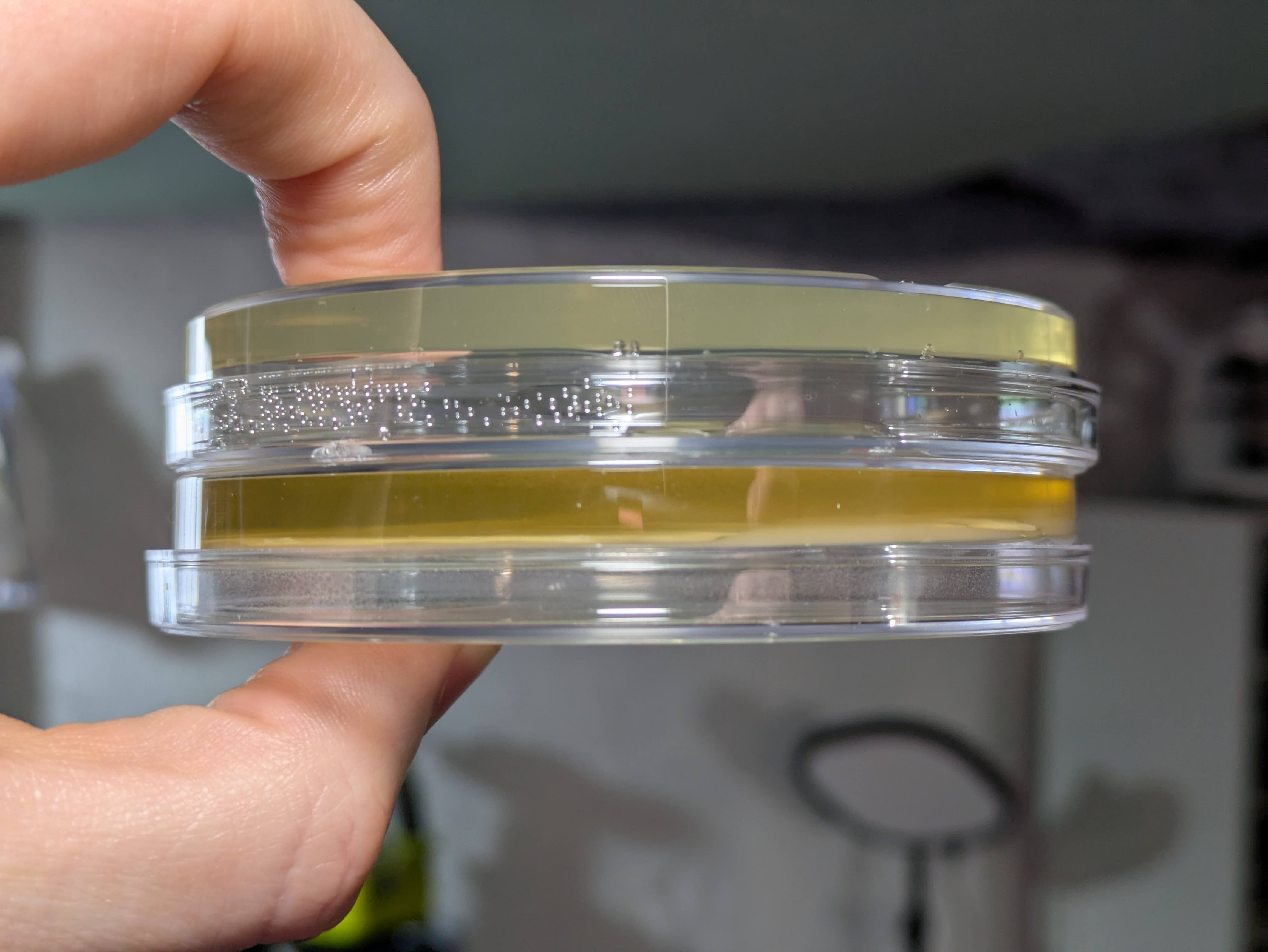
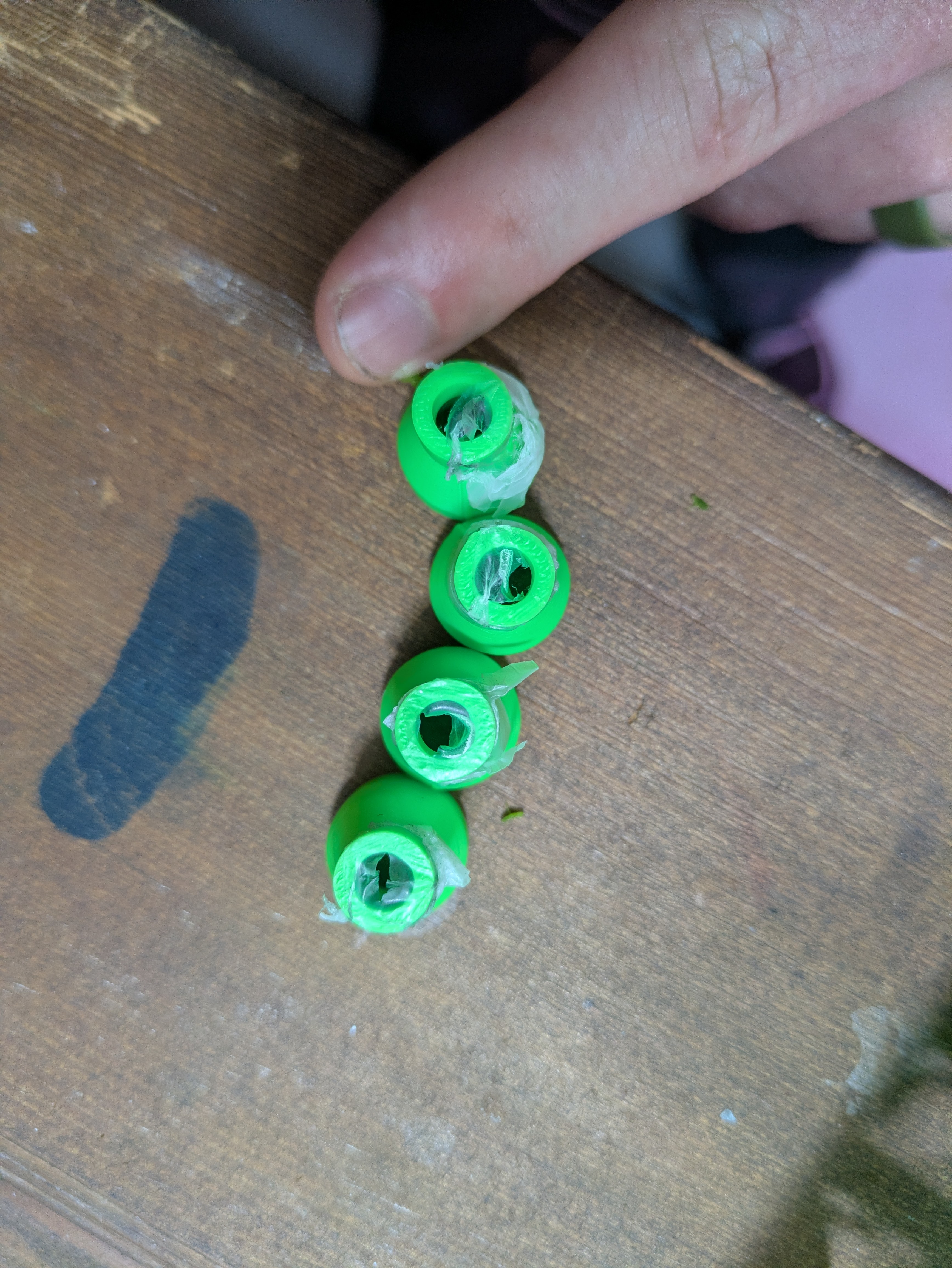
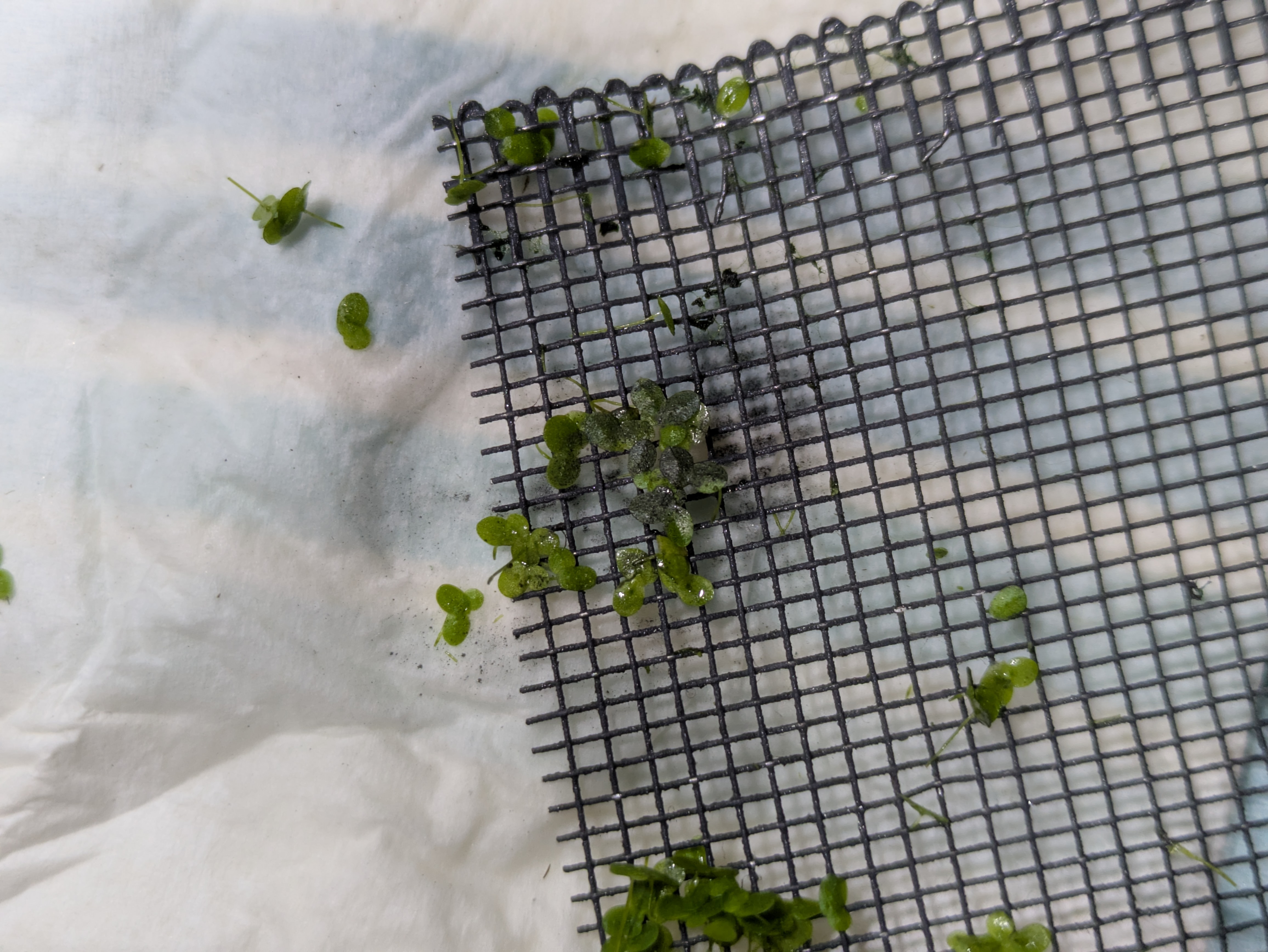
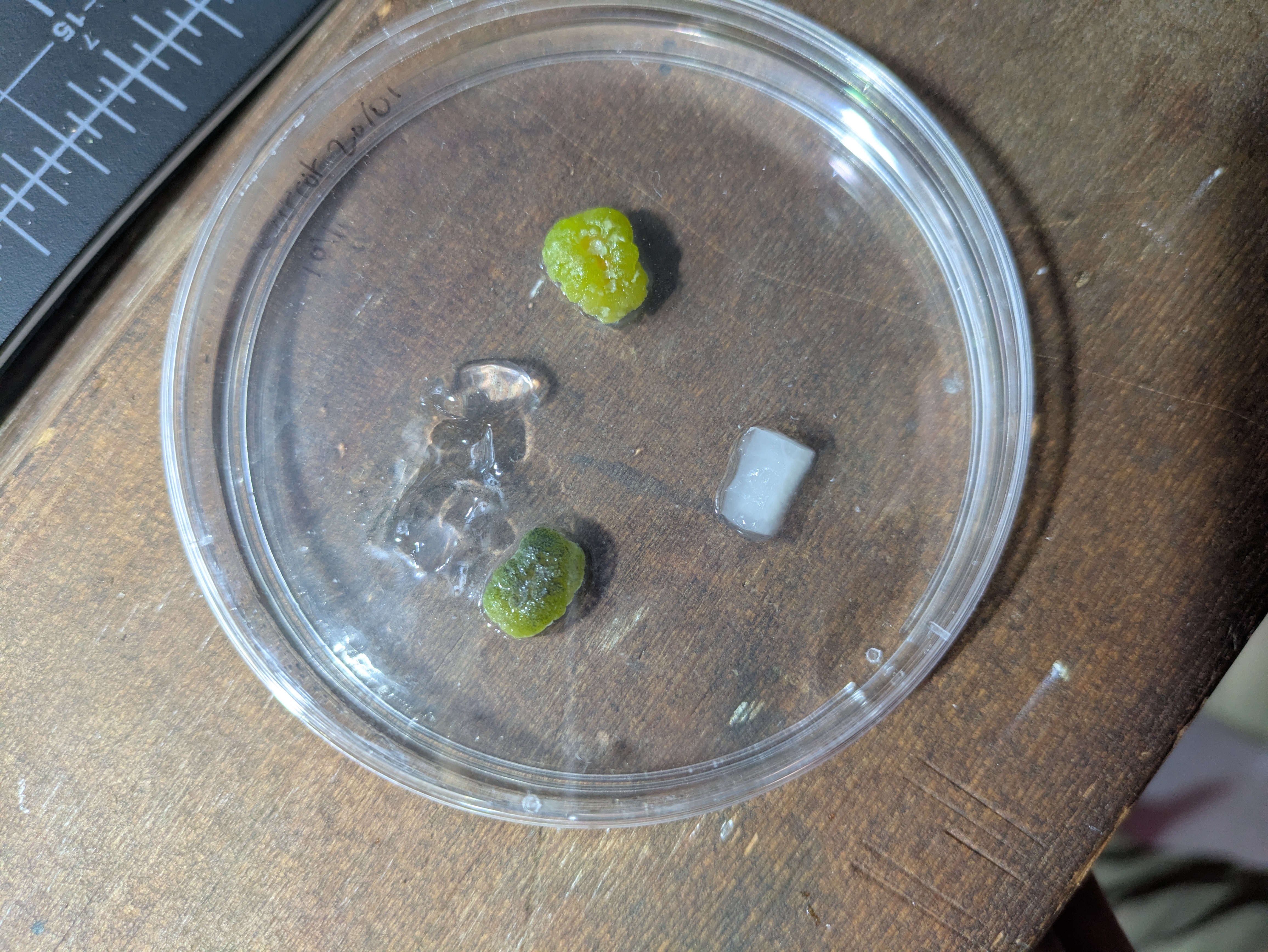
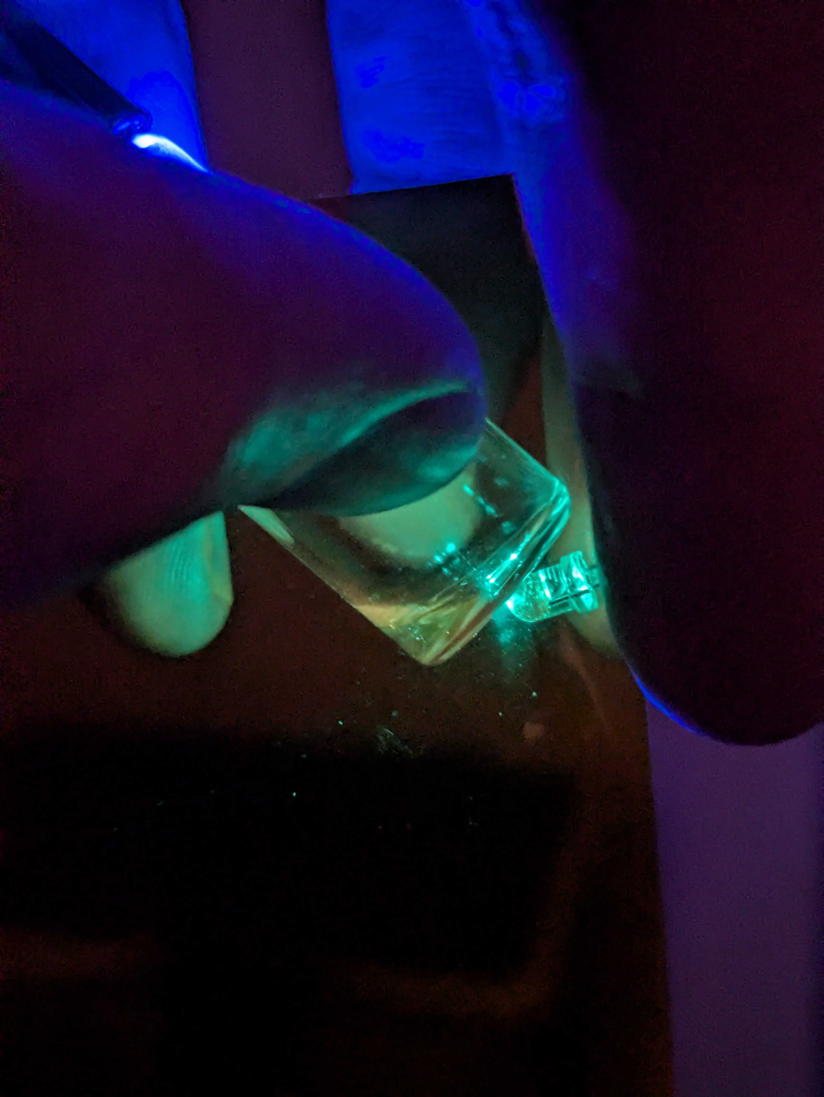
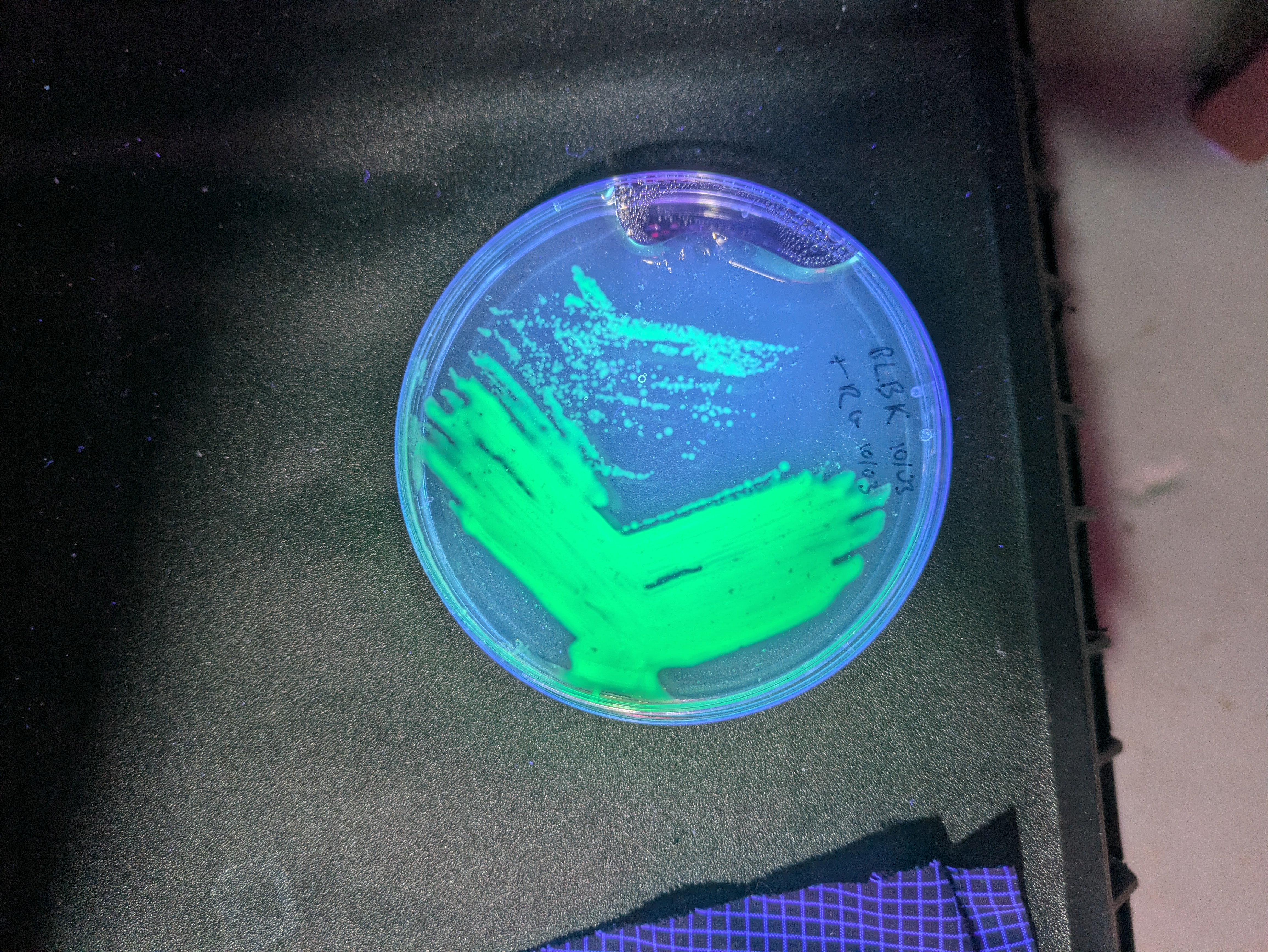
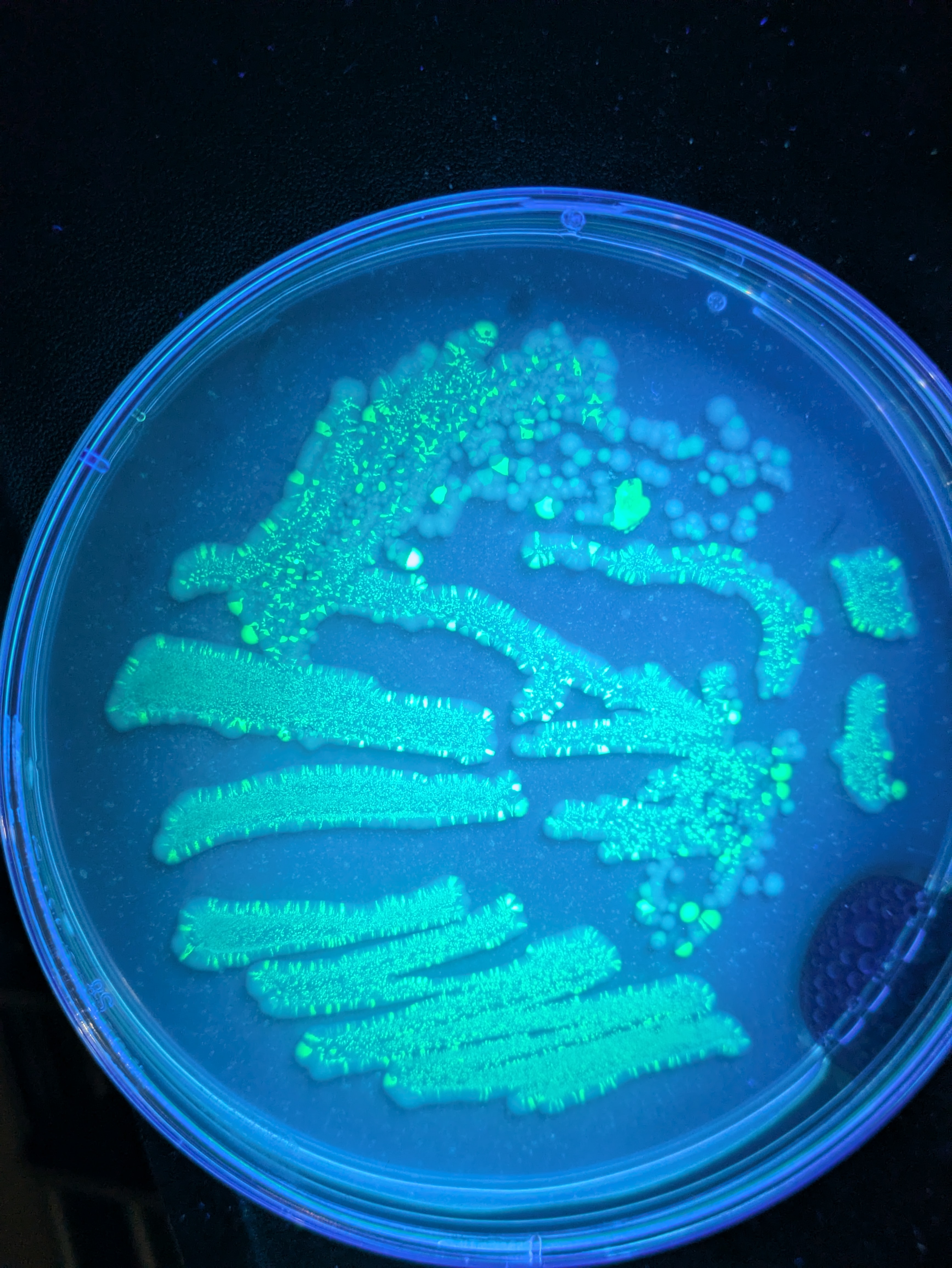
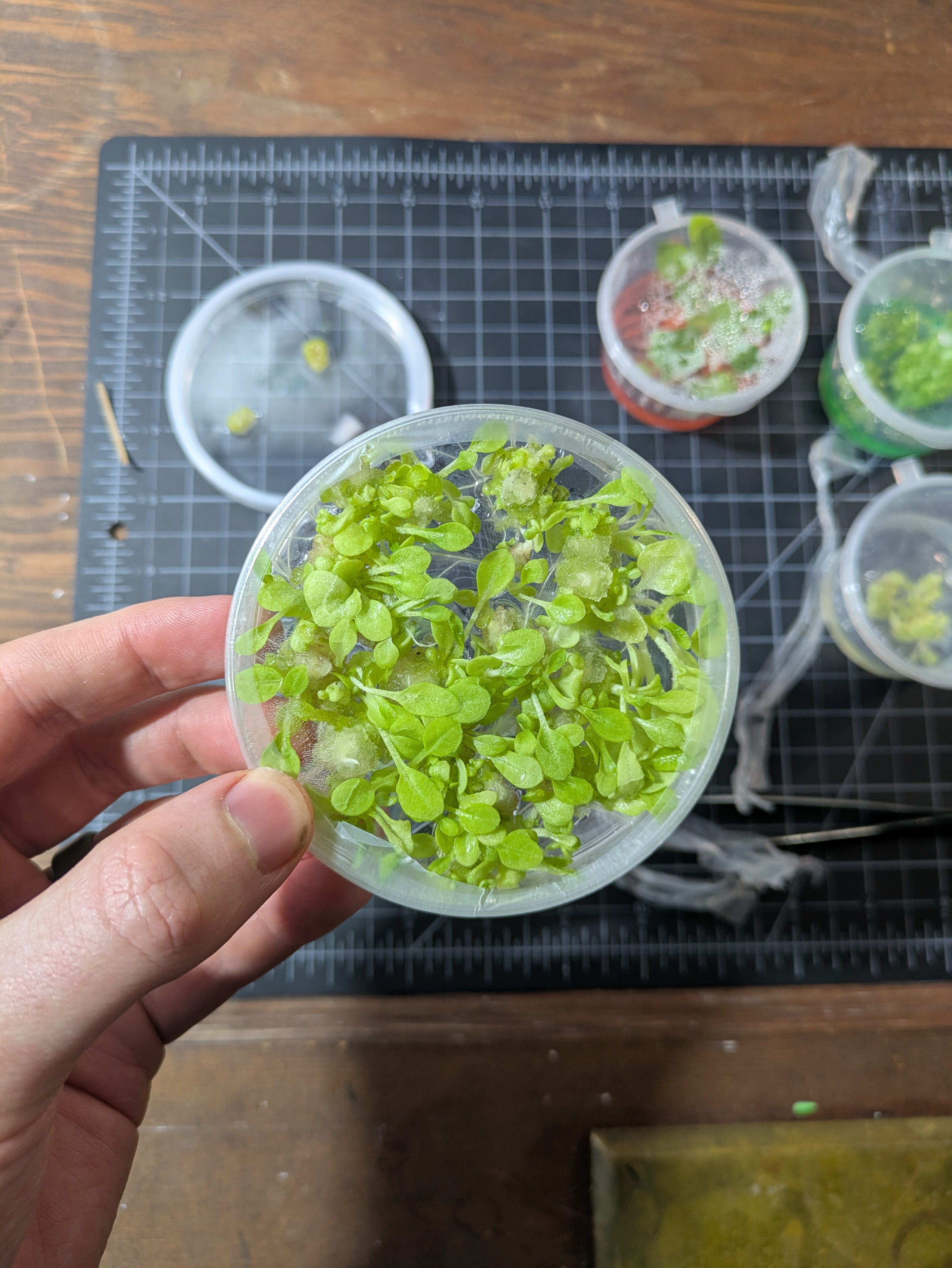
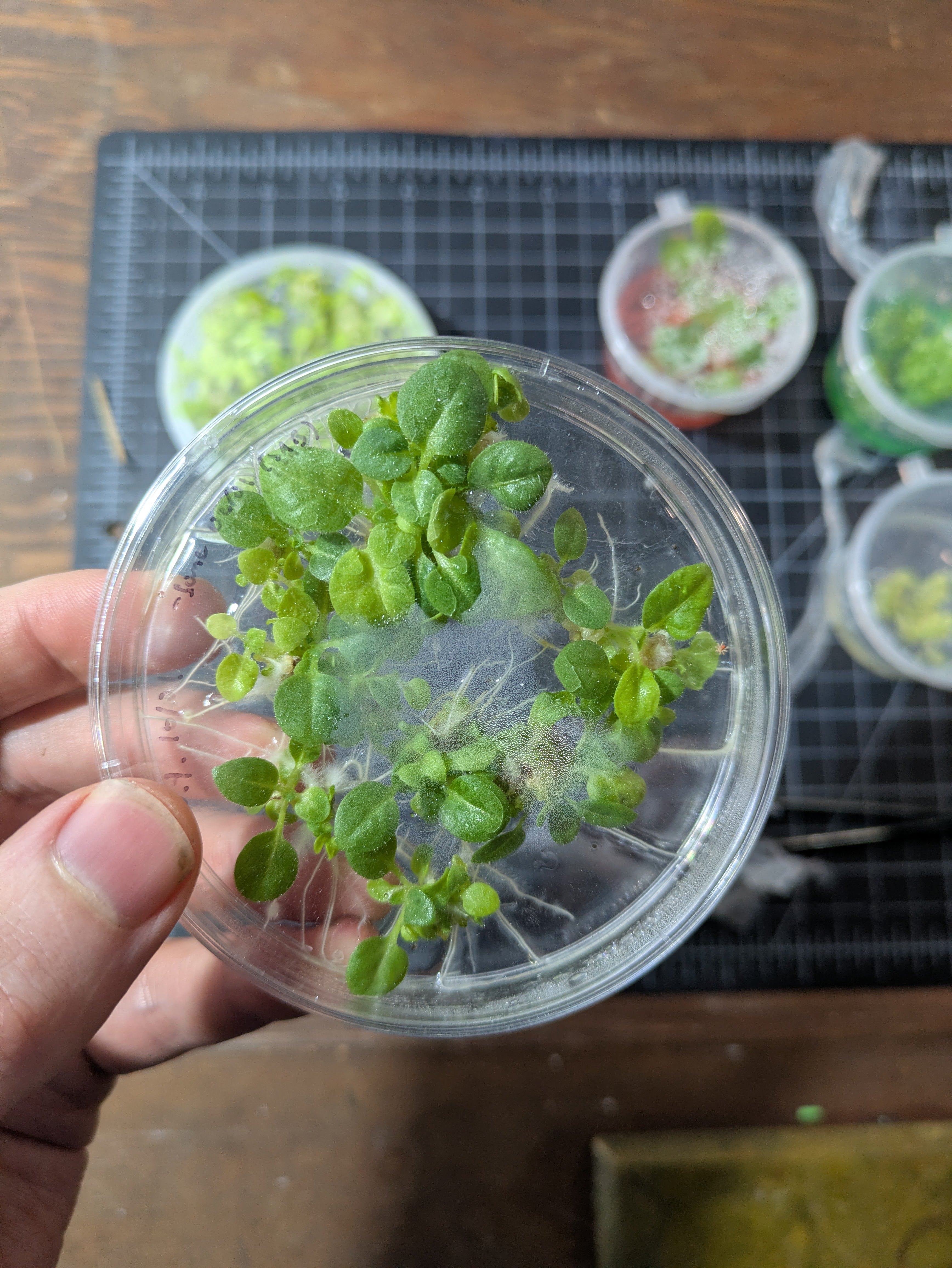
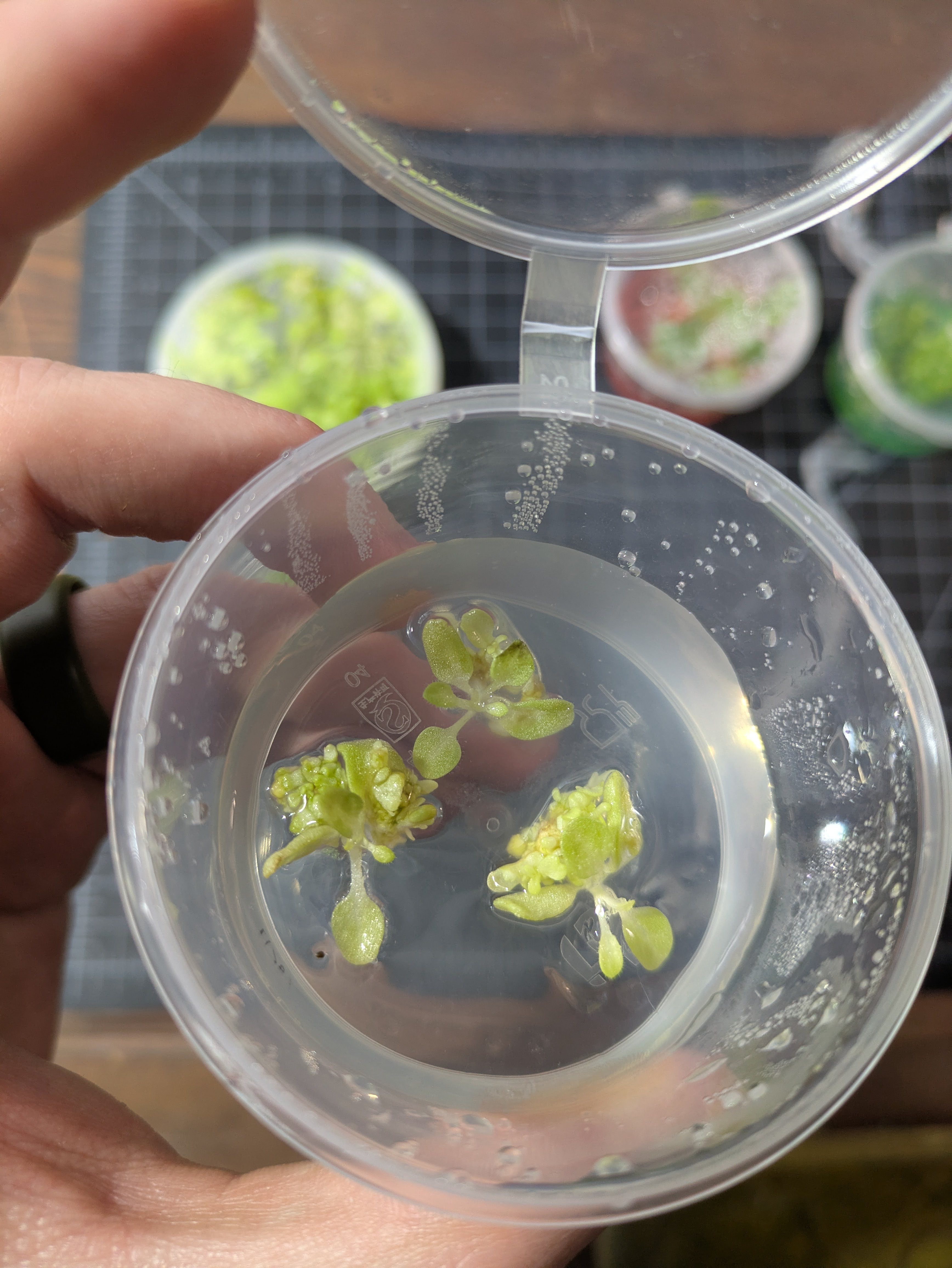
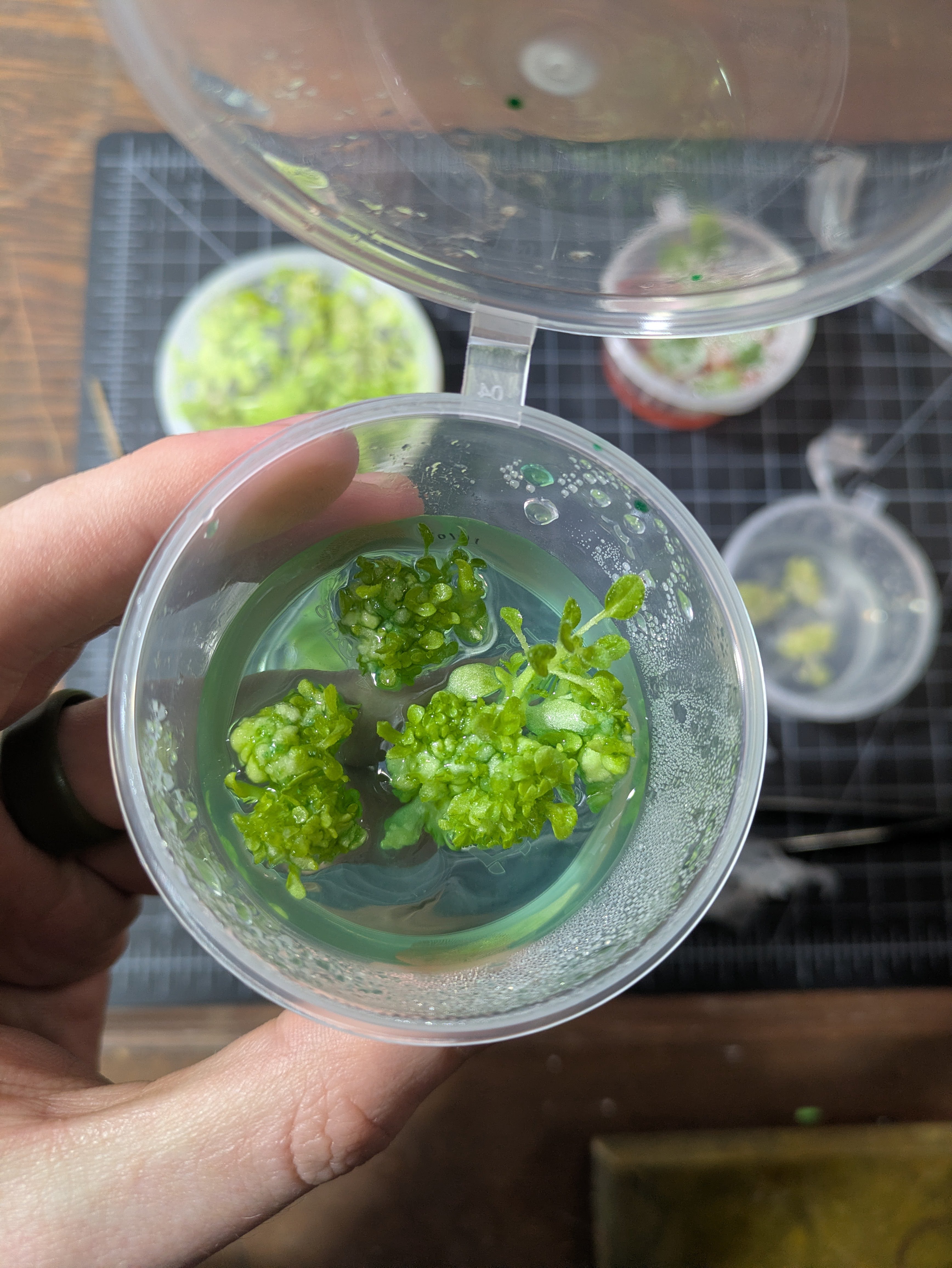
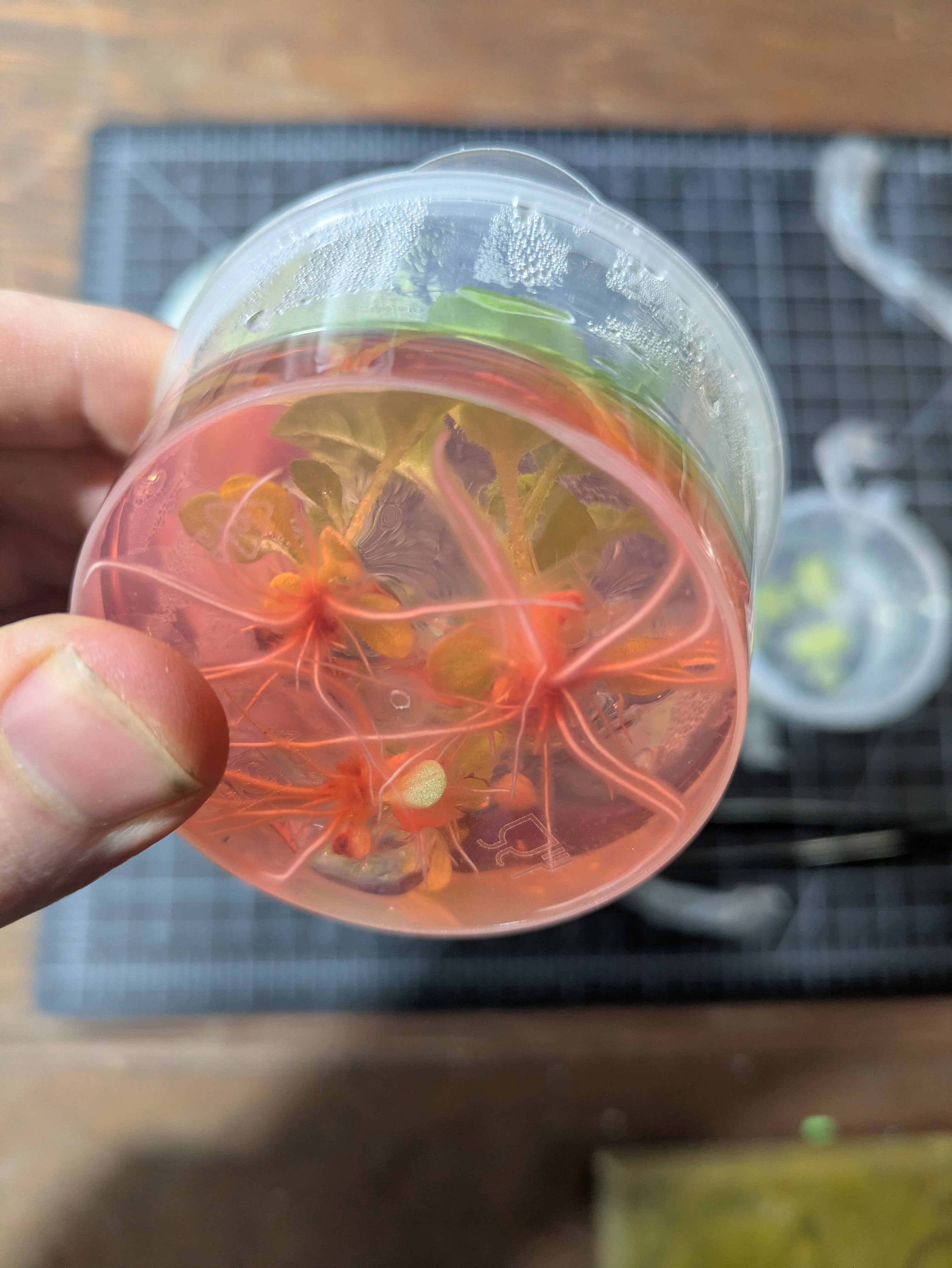
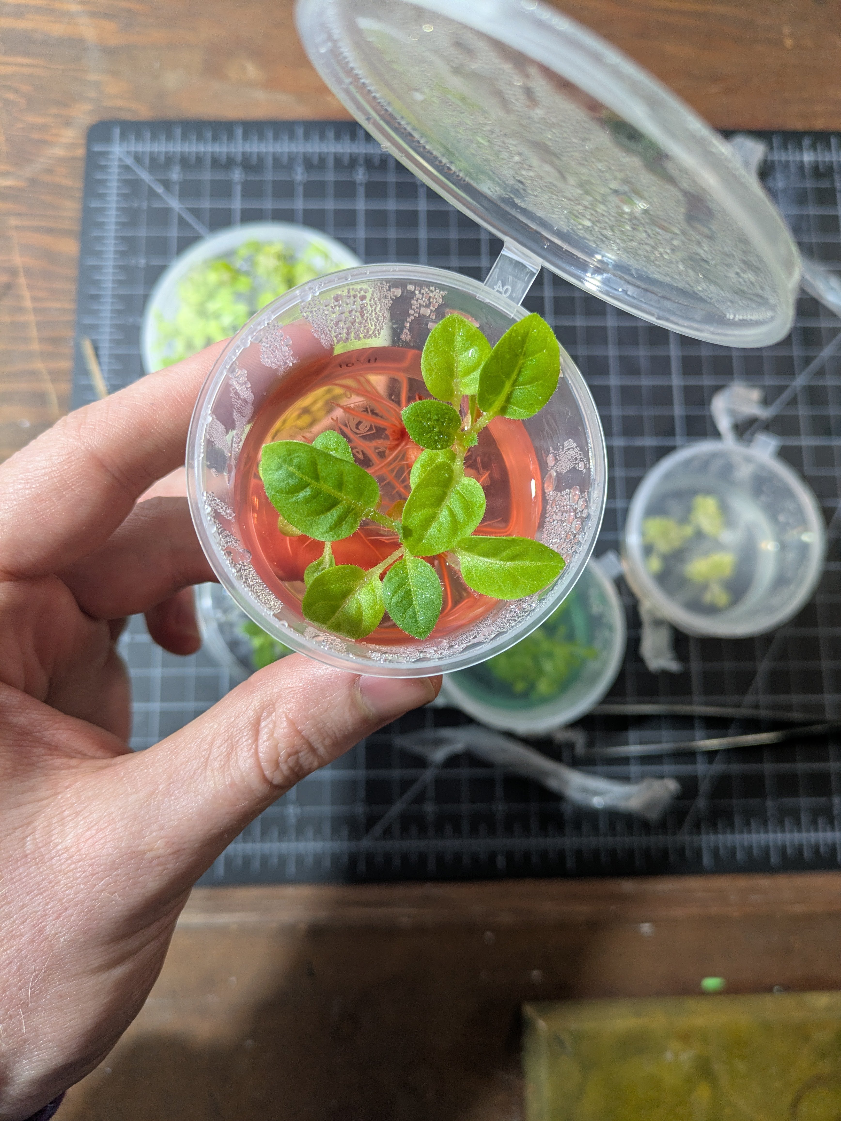
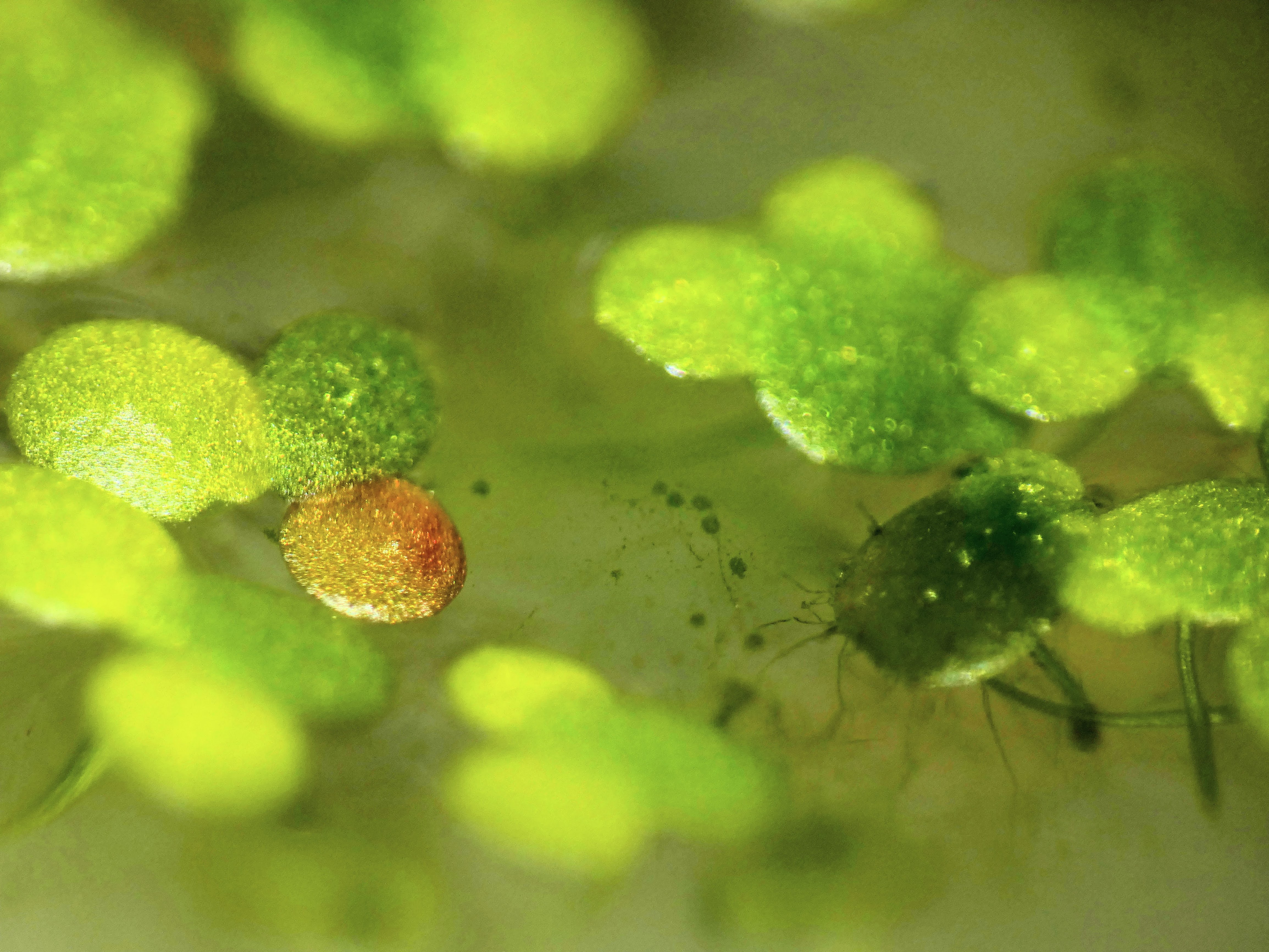
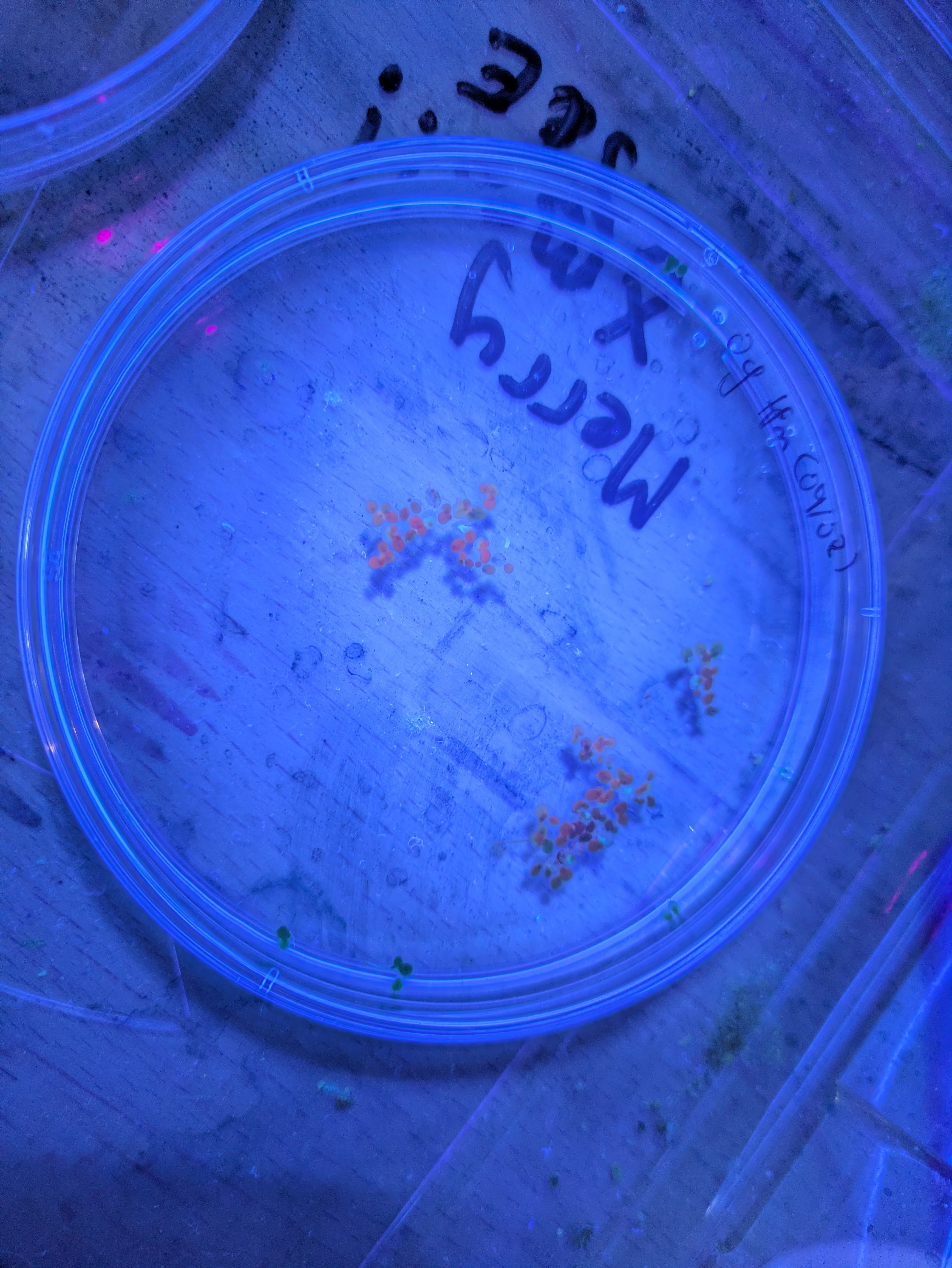
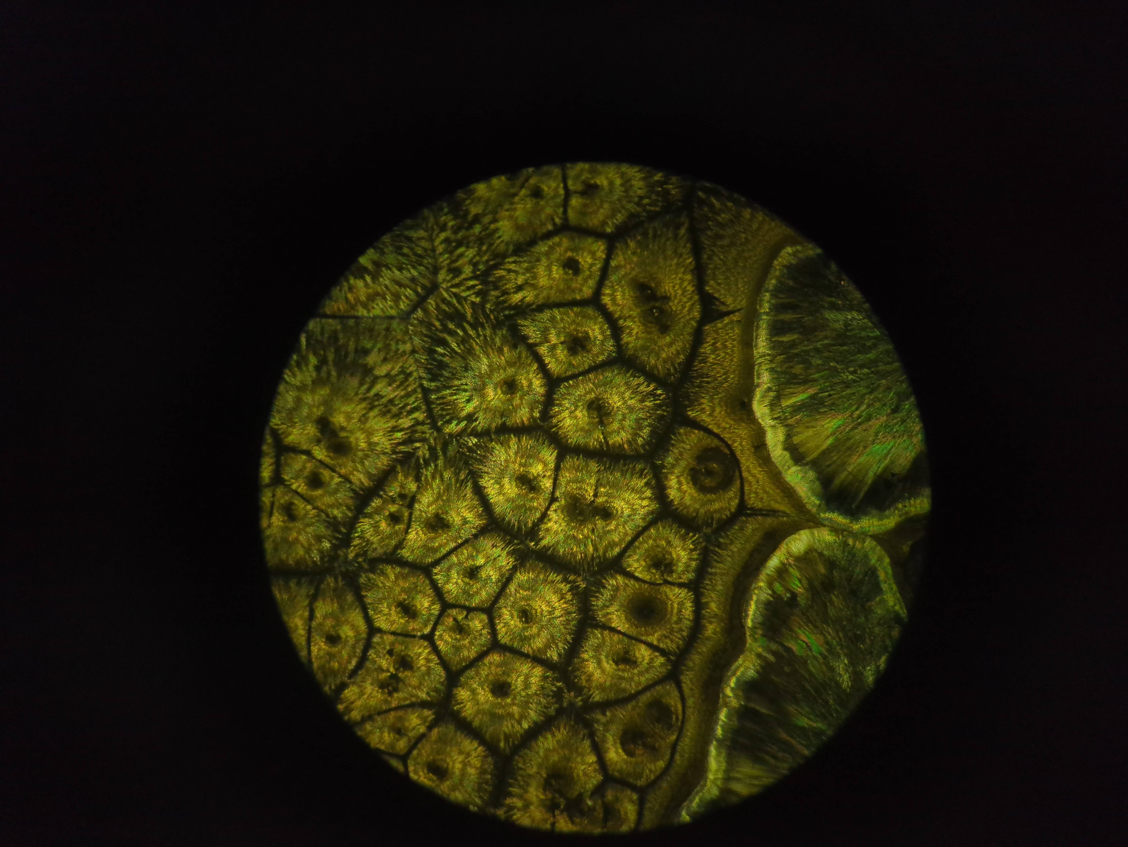
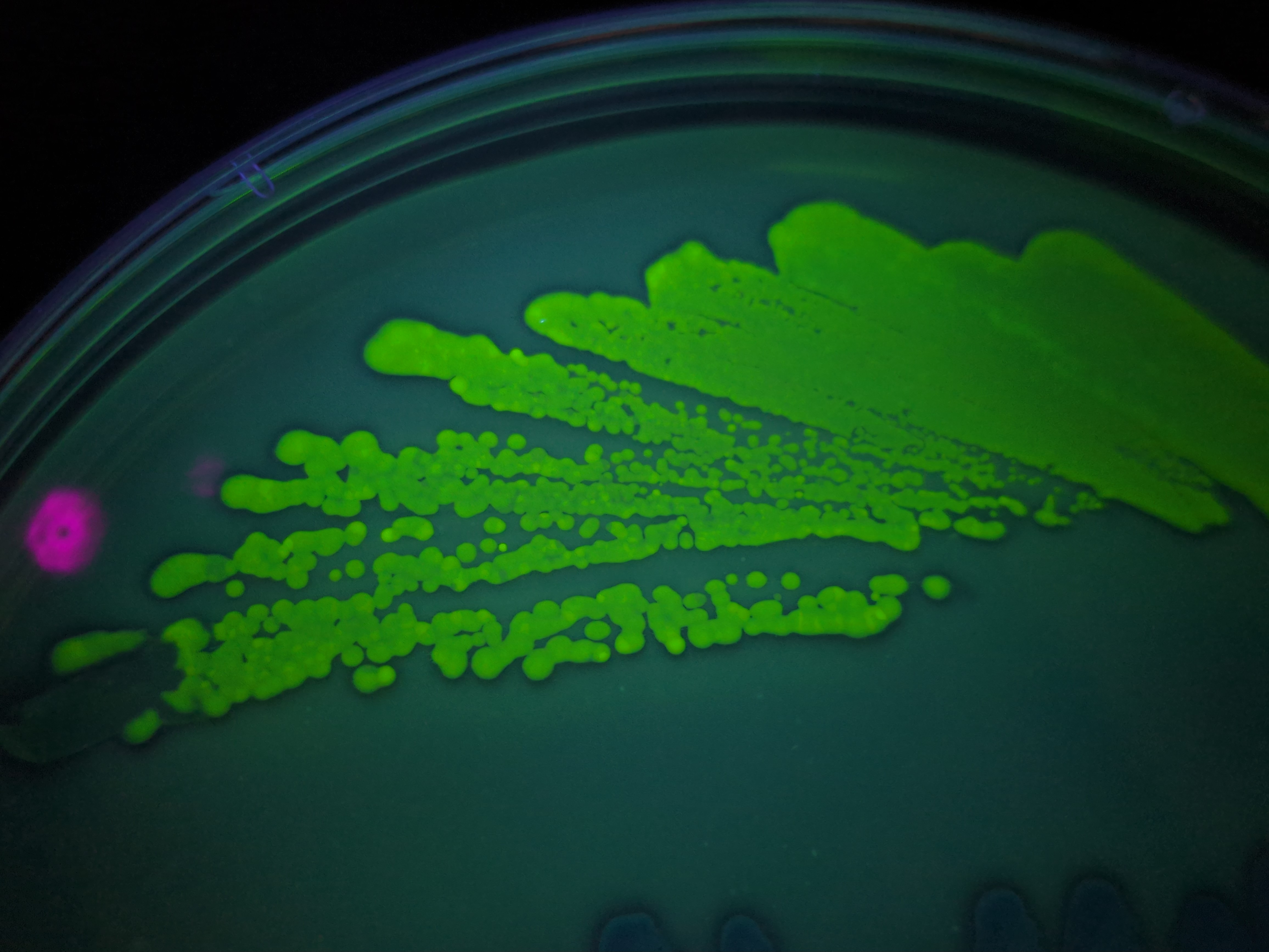
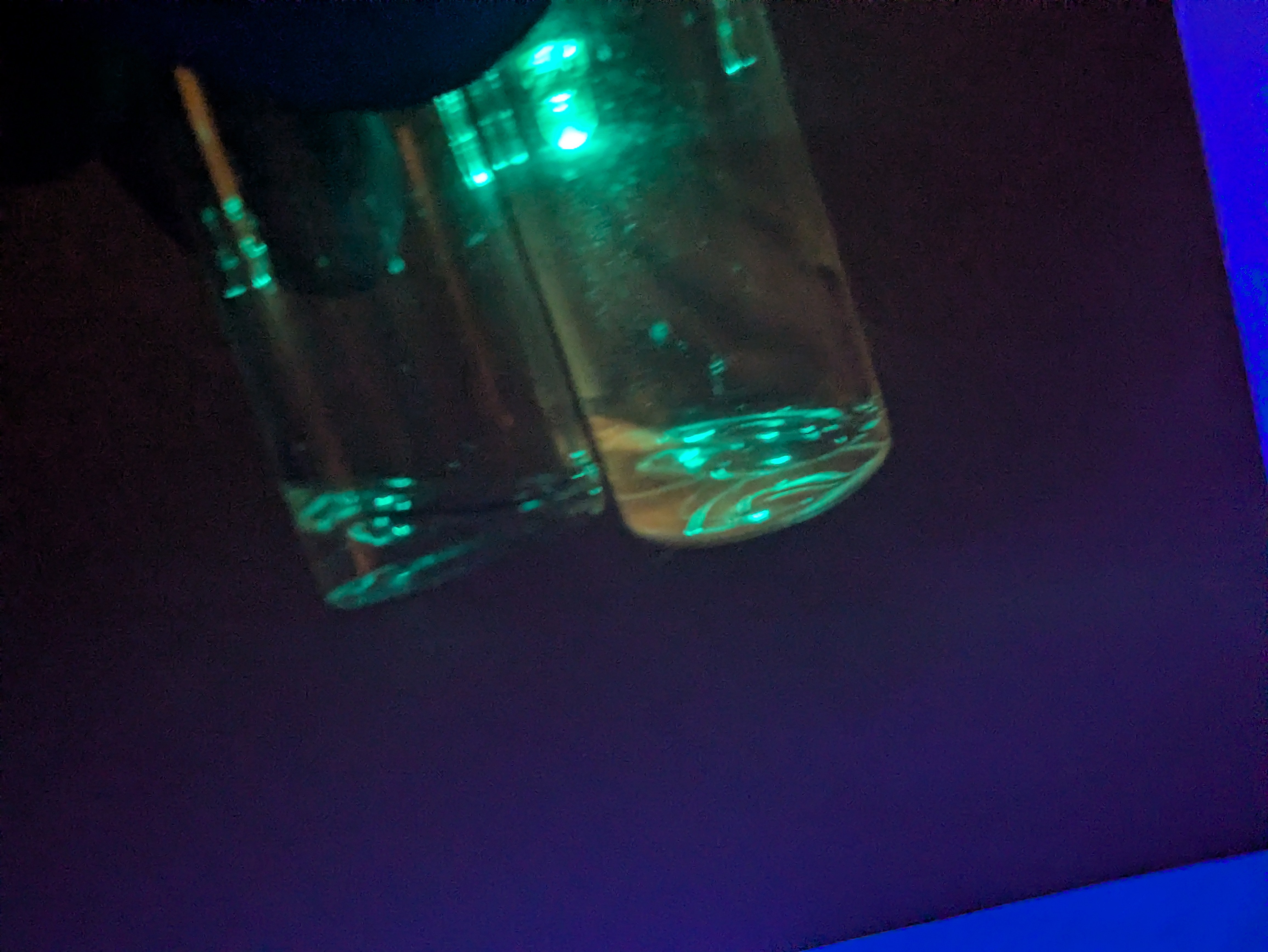
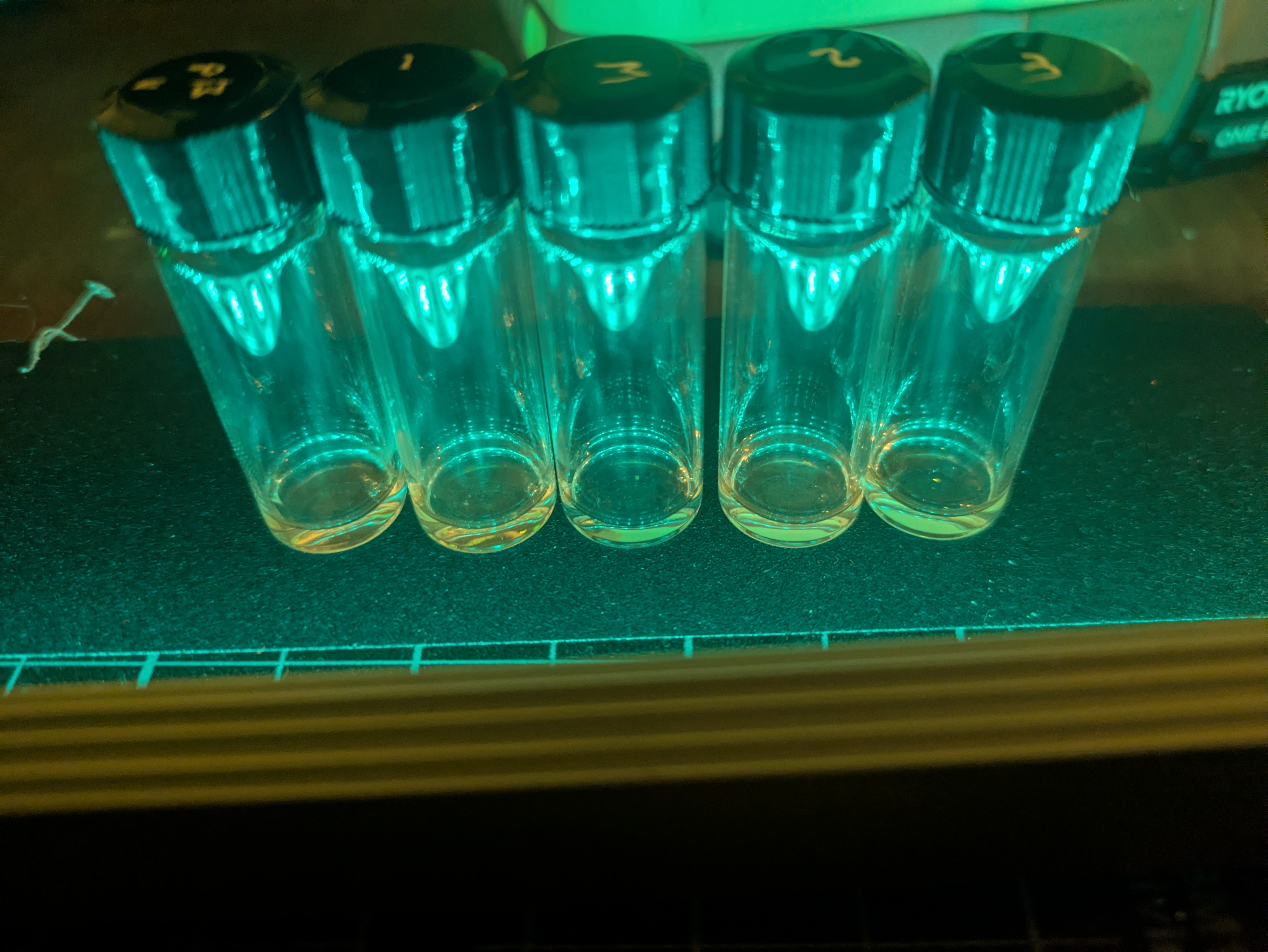
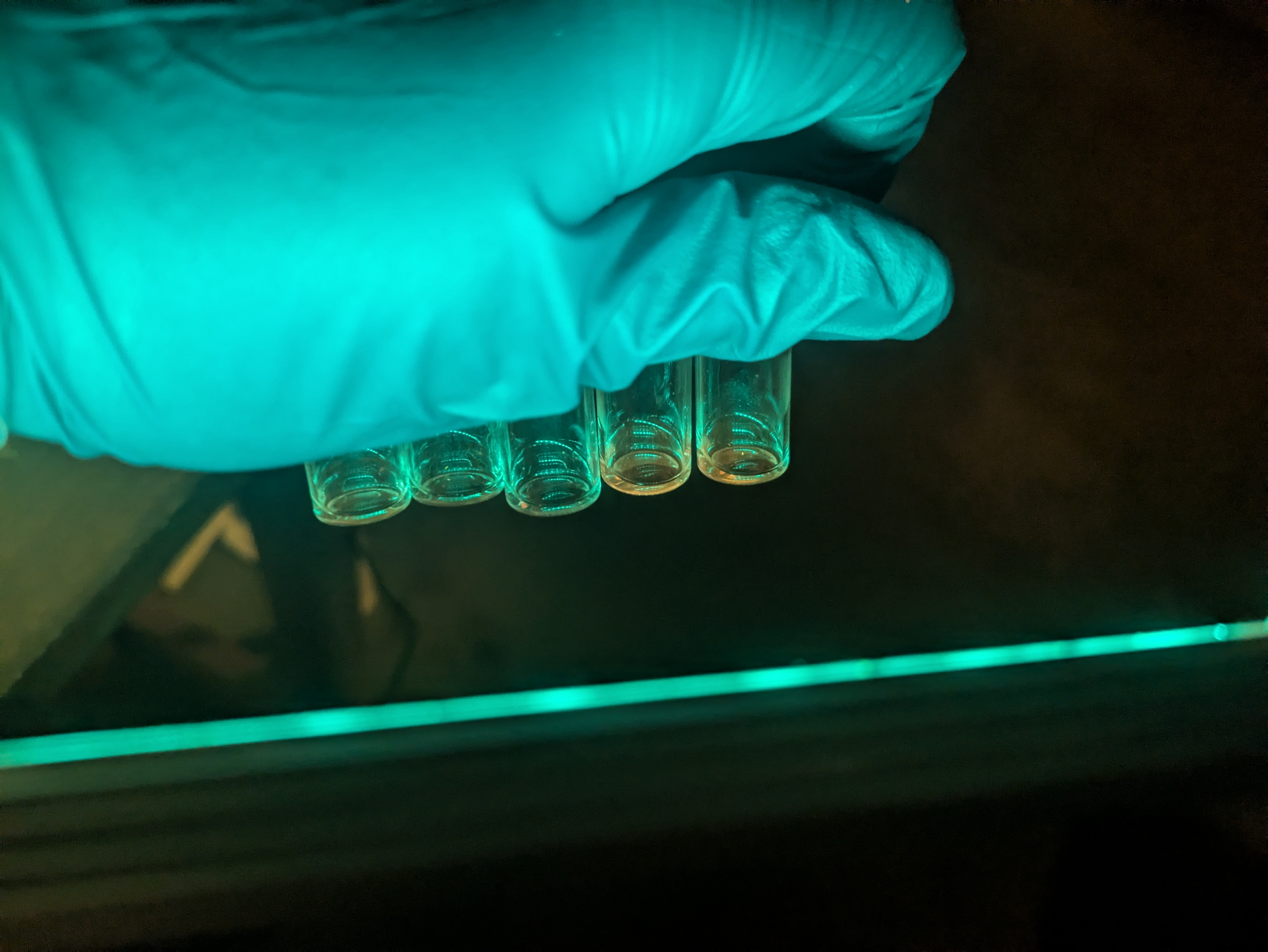
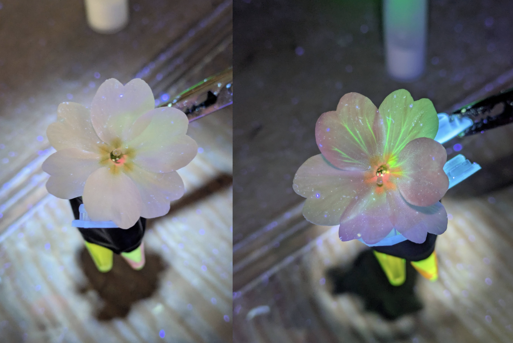
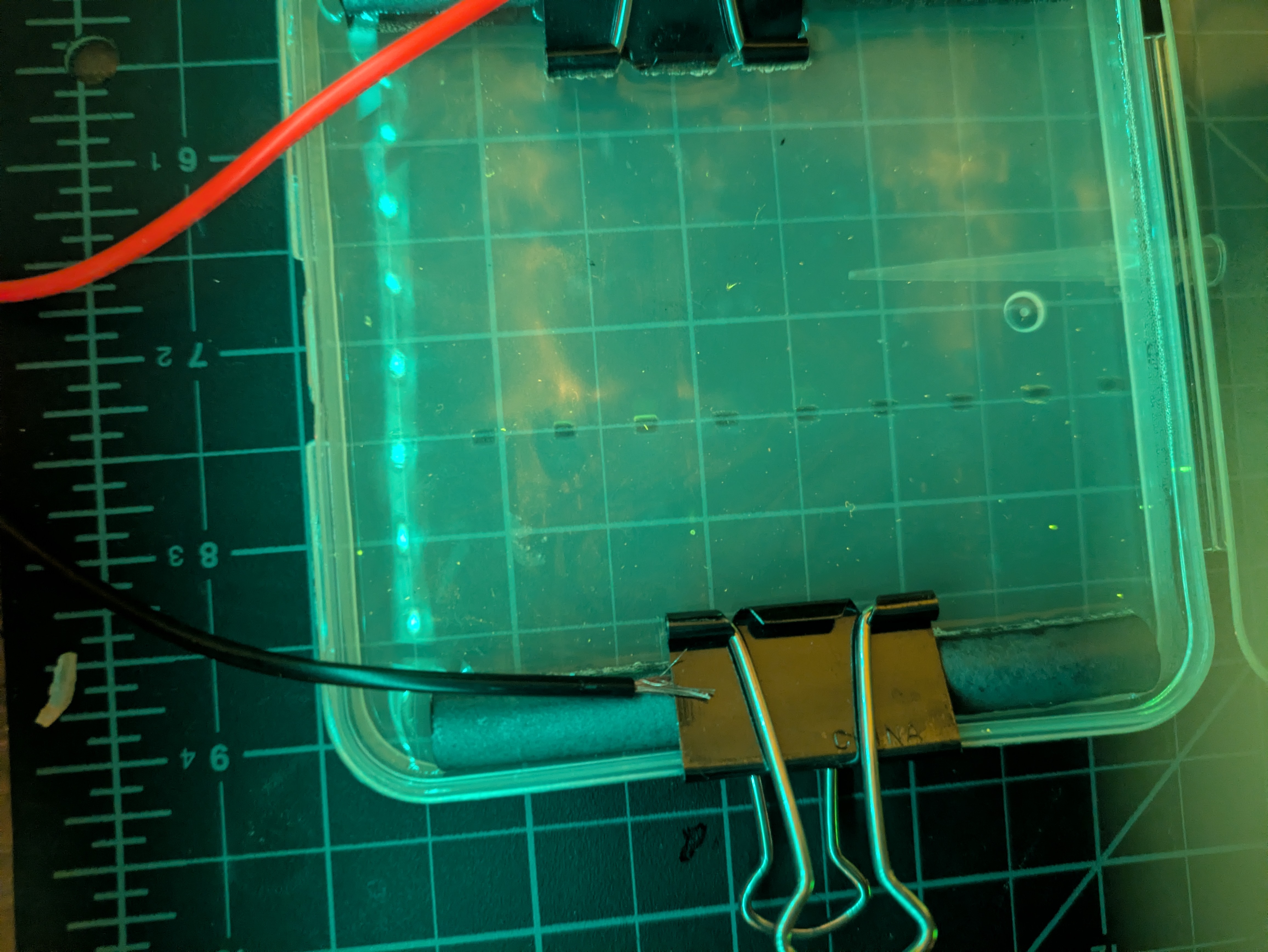
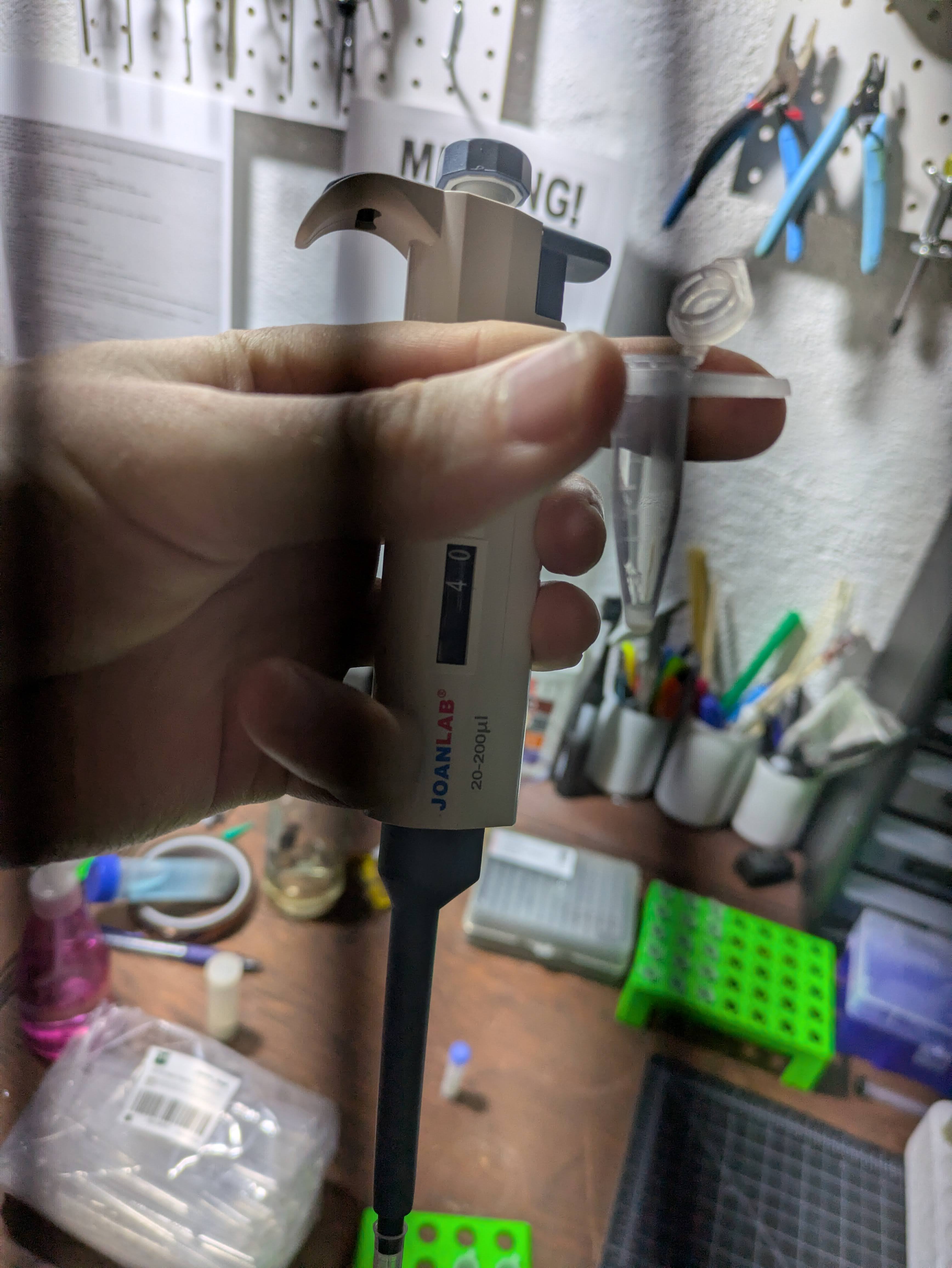
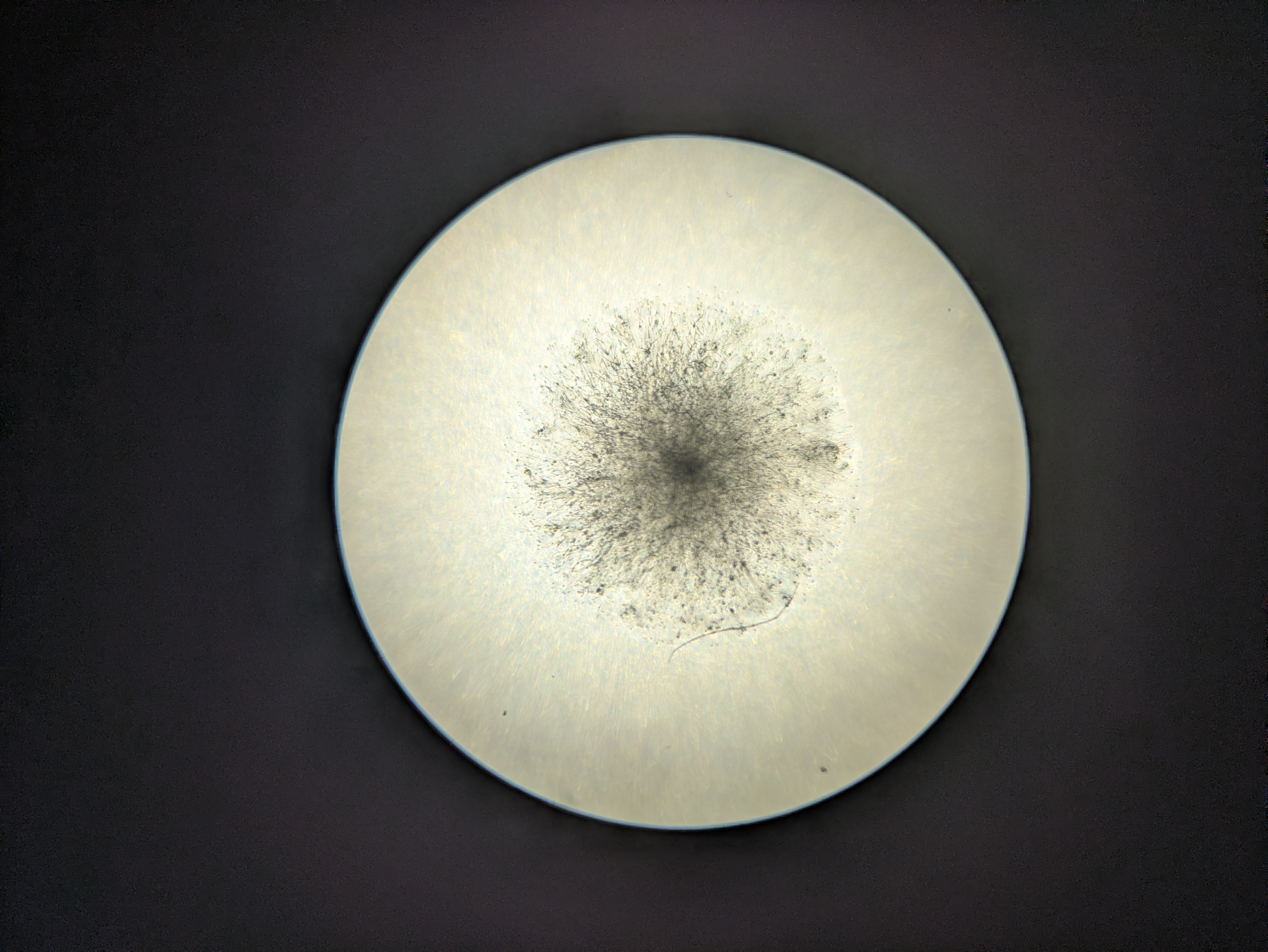
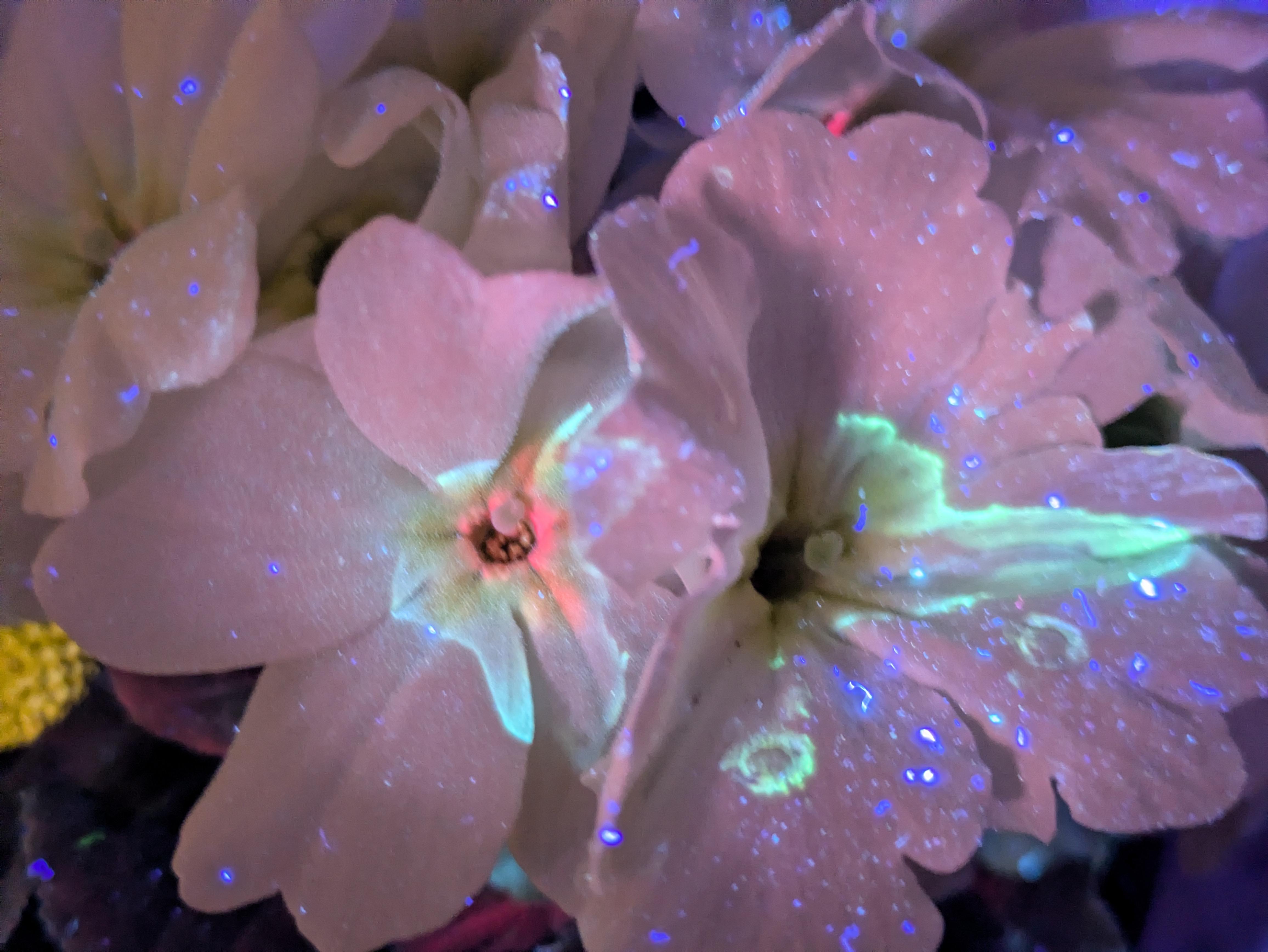
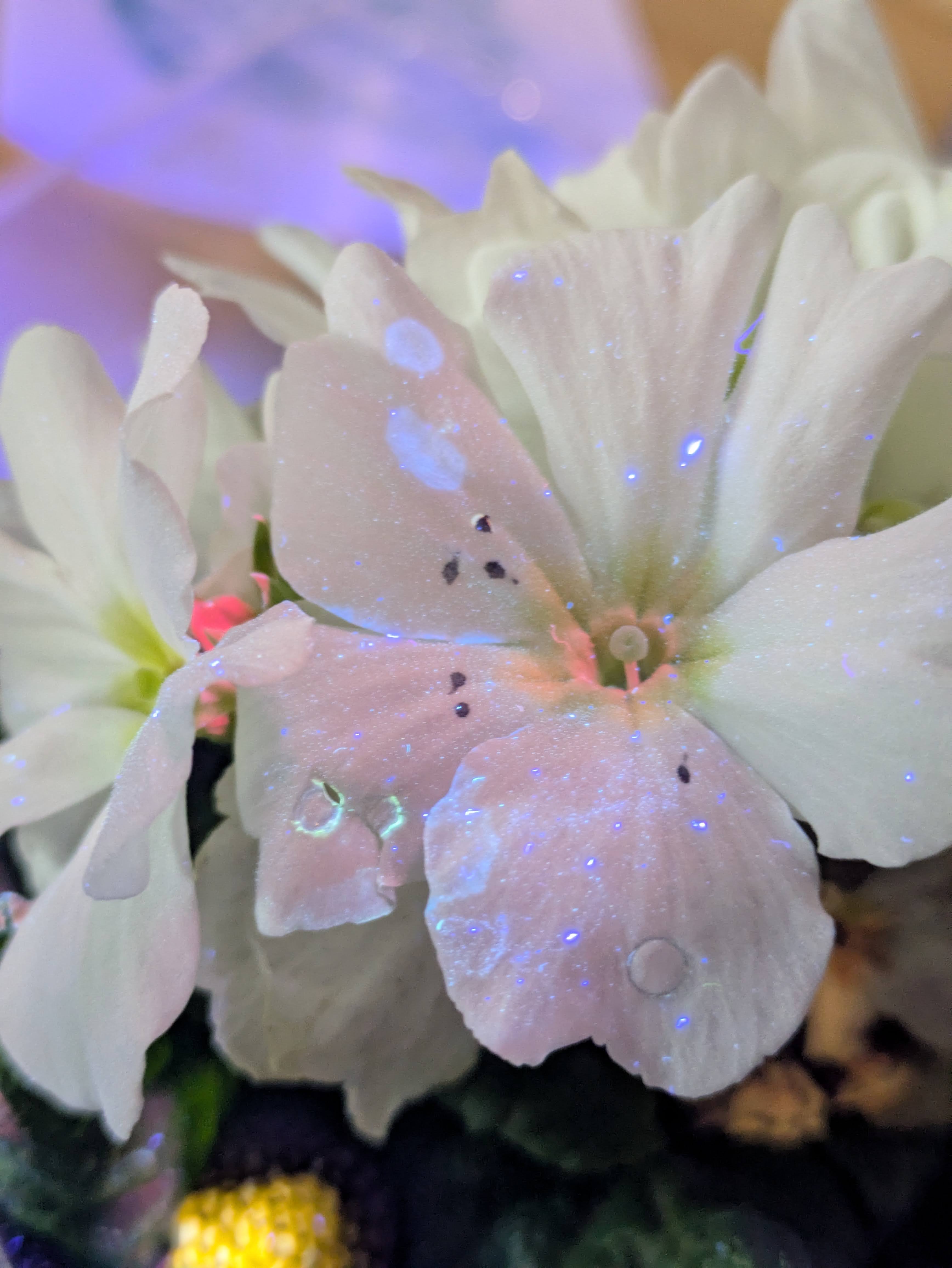
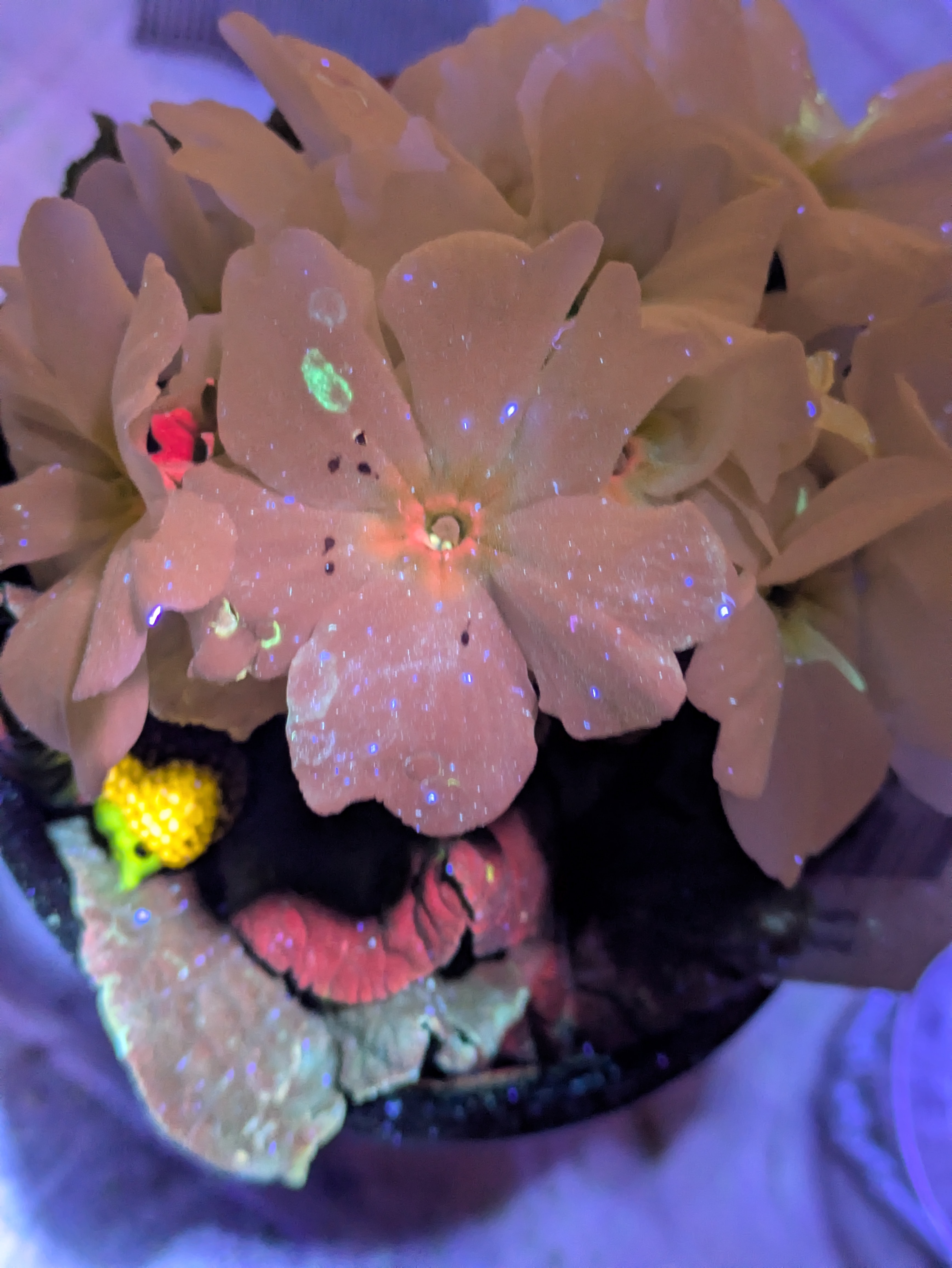
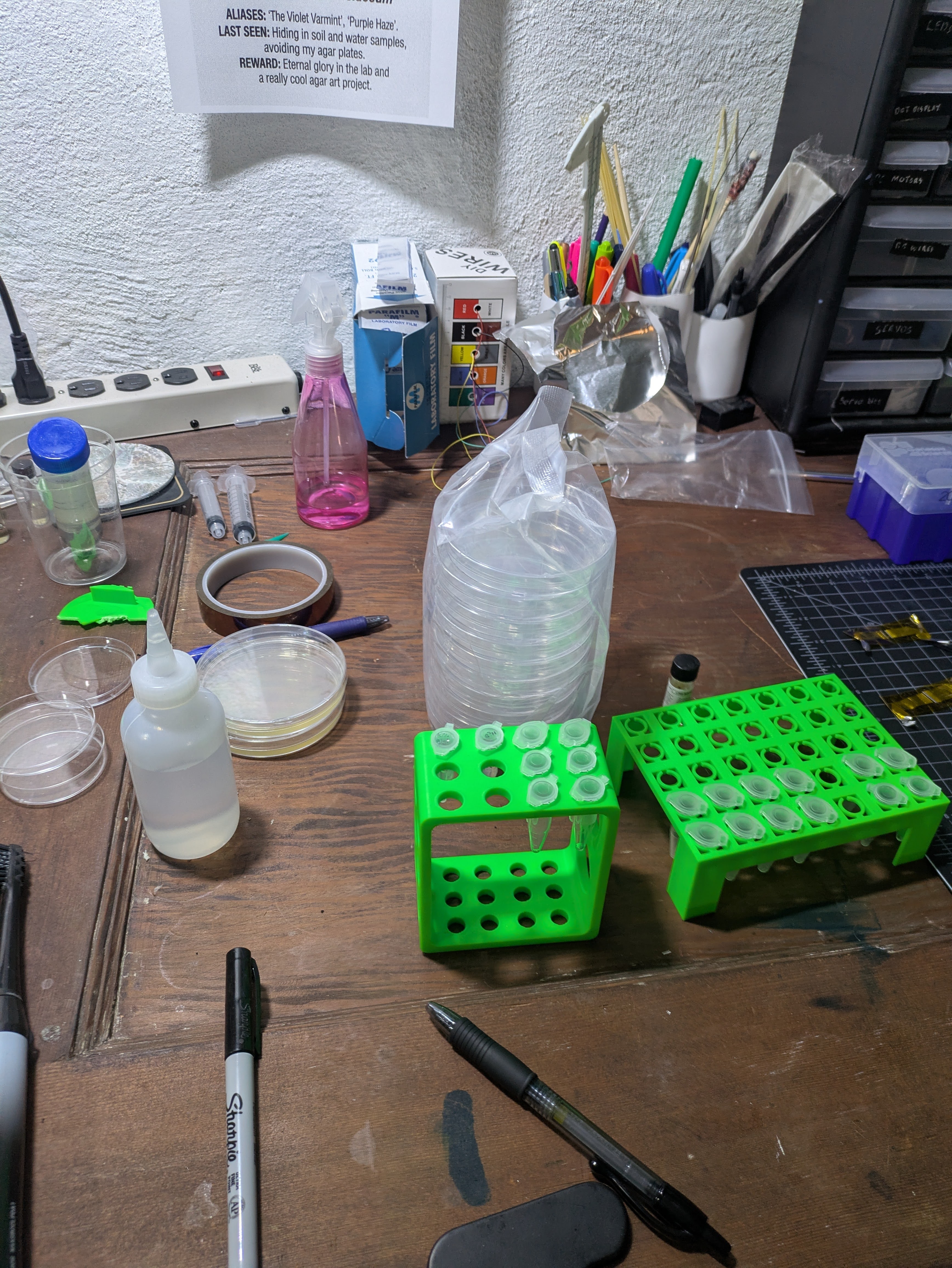
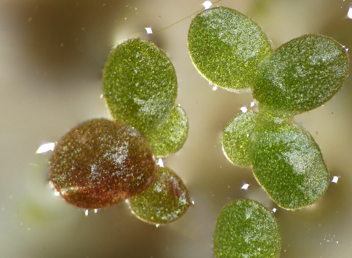
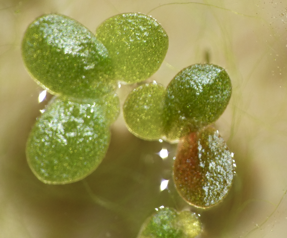
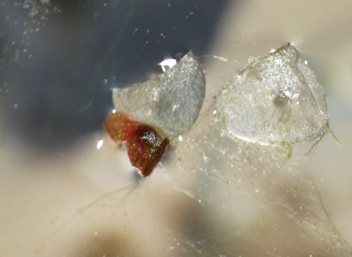
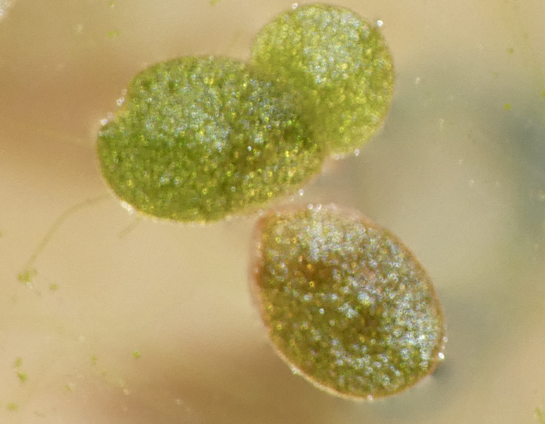
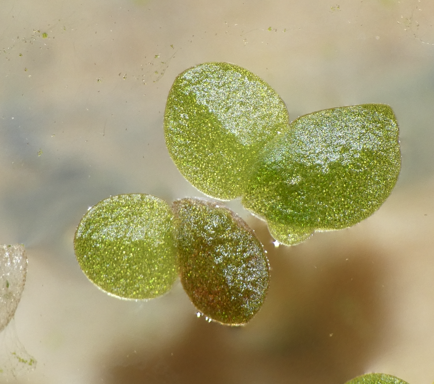
More pics of duckweed with RUBY, transformed with agrobacterium. I still need to try a fresh attempt, planning to split them in hopes of transforming more meristems, but even the rough and ready way I did these at least yields some partial transforms and a couple of fully transformed bits! Hopefully over successive generations we can weed out the mosaic mixes and get pure, but if I can up the initial transformation efficiency that'll help. How these were done:
- Agro + tfm mix in a syringe
- Add some dwe fronds
- Pull back and release plunger a few times
- Rinse a few times
- Into nutrient mix to recover + grow
- Wait.

Ruby???

Permalink

Inspecting the leaf I shot with DE + CaCl2 + RUBY DNA has a few tiny red spots, hopeful???

Biliverdin diffusion

Permalink

Well, I tried growing out the e.c. with my biliverdin plasmid. No obvious color in the colonies (maybe a little more yellowish than normal?) but the agar over time has shifted color. I think as I feared the biliverdin is diffusing out, and it's more bruise-brown than obvious green in the LB agar. Might need to try operation treefrog.
Also, this is growing out the cloning strain I got from genscript. The plasmid DNA tubes they sent looked empty, with the dry ice I suspect the DNA was freeze-dried in the (no parafilm) tubes. I added sterile water, and tried out my spec method to see if DNA was present, seems like probably? I did a transform but got one (1) colony so something funky going on. Next trying liquid culture of the stuff I already have, and leaving things for longer to see what happens.

More gg tests

Permalink

Trying 10ul slurry, 10ul CaCl2 soln, 2ul DNA, letting dry, with both de slurry and graphite. Tried into tobacco leaf and also duckweed

Misc

Permalink
  • Gene gun tests with graphite into leaves -> no ruby I can see, killed a lot of the leaf in the center of the blast. Tungsten needle tatoo vibe also killed the tissue. In onion, I got a couple of ambiguous reddish bits, not sure if TFM and RUBY expressing or something else.

I got my biliverdin plasmid delivered today, TIL if you pay $10 extra for glycerol stock you get a buttload of dry ice with it, made a janky cloud chamber and watched some rays, had fun with the baby playing with bubbles, blew the lid off a tub a few times :D

Got a taser but at 25kV it's a bit too powerful for electroporation, need to come up with a plan for that.

Pouring some plates and making some stabs now.

Quantifying DNA (test)

Permalink

Shining a blue LED into 100uL of
- SeeGreen DNA stain (2ul in 20ml)
- with 1uL of soln of interest

Seems like uL (blank), w (water), 2 (an old agro miniprep attempt) have very little. spB is an aeBlue mini-prep test, s3 was my final pB practice run, and uR is my RubyGreen mini-prep that actually matters. s1 is 10ng/uL (too weak to see) and s500 is 500ng/uL approx (maybe some tube losses, could be 300 for all I know) of pBLY. Not at all calibrated, SNR is terrible, but I sliced it a few different ways and re-tested and uR always shows the biggest peak, don't think it's too far-fetched to say 30-70ug of DNA in the 100uL elution I managed to get! (in the mini-prep I used a big loopful of cells, filtered the stuff then ran 2 ~800uL portions of the filtrate through the column to maximise my chances, and eluted with 70uL then an extra 30uL (letting the latter sit a minute) to eek out as much as possible.

Edit: re-ran more carefully, est DNA conc 687 ng/µL (90% CI: 507–845), code here: https://gist.github.com/johnowhitaker/3aa41553b027c29d307766fcc06a79cf

Shooting DNA at things

Permalink

Trying graphite (powdered on 220 grit sandpaper, 200ul loose powder volume in 1ml water and mixed up as a slurry, 10ul as a drop allowed to dry) as a carrier, with 2ul DNA from the miniprep (dropped on then also allowed to dry). Shot various things plus a few no-dna control, yolo run but let's hope we get at least a glimmer of ruby in a few days time! For now, pie party. My melktert recipe:
Into a saucepan put:
- 3 Tbsp corn starch
- 2 Tbsp flour
- 1/2 cup sugar
- pinch of salt
- 1 tsp cinnamon
- 1 Egg
- (mix), (splash of milk), (mix), 4 cups milk
- (heat and whisk till thick and custardy, 5-10 mins)
- knob of butter, vanilla (1tsp)
Pour into graham cracker base from the store, dust with more cinnamon, cool to room temp, set in fridge a few hours.

Rg miniprep

Permalink

Miniprep on pCambria RubyGreen, think I got some DNA? Todo machine to quantify amt. Also planted plants a few days ago.
My 'fuge is weak so in the miniprep I use a syringe filter rather than spinning down after mixing all the things, and just give it extra time when sending stuff through the column. Also for this one I just scraped a big loopful of cells from the plate into A1 rather than growing out liquid culture

Tobacco TC tests

Permalink

Cracking open some tobacco seed to TC tests I started in mid Jan. 3 diff media formulations.
Base media: 400ml water, 2g MS, 3.5g agar, 12g sugar. 25 ml / tub
Green: 25ug BAP, 3ug NAA (in 25ml)
Red: 6ug NAA
Clear: 25ug BAP, 6ug NAA
Seeds plated on clear, 3/tub transferred after a month or so.

Duckweed tfm

Permalink
Notebook page for 2026-03-03

Attempting tissue-culture-free agro transform of duckweed (lemna minor). I ran a few diff exps this weekend, and more excitingly noticed a red frond in some that I'd tried to tfm on Feb 9th, hoping that means the meristem took in the genes and this is a properly transgenic new frond. Rough protocol:
- A loopful of agro into infiltration mix, in a syringe with some duckweed fronds
- Pull back plunger while blocking end, and release. The sudden vacuum+slam (in my head at least) helps push agro into the interstices of the fronds.
Will share more depending on how more recent tests go.

Misc

Permalink
  • a couple of colonies visible by this evening on the electroporation plate(s?). Will evaluate tomorrow.
  • went down the rabbit hole of DVD OPU microscopy options, AFMs, flexures etc again
  • modelled and am printing bits for a nicer spectrometer

Electroporation take 3

Permalink

New run:
- Pseudomonas in liquid culture for ~18h
- Spun down, removed supernatant, add 10% gly (in 1.5ml tube)
- Spun down, removed supernatant, add 10% gly
- Spun down, removed supernatant, add 30uL of 10% gly (pellet by this stage was pretty small given my weak 'fuge)
- Added 3uL DNA (from a mini-prep I did yday, aeBLUE on a kanamycin resistant backbone)
- Did this in two tubes. One got 1.2mm gap, one got 2.4mm. 10 zaps each. On the 2.4mm gap, peak was ~5V (~10kV/cm). On the 1.2mm gap I didn't get a clear voltage reading, not sure what went wrong.
- 1h in 200uL LB broth @30C to recover before plating 100uL on Kan plates. Now we wait. Only worry: cells might have been stationary phase, might try a repeat with fresh culture in a few hours.

DNA after all?

Permalink

I ran another mini-prep, although since my centrifuge is funky I used a syringe filter after the precipitation step. Then to see if there is DNA there, I added 2uL SeeGreen stain to 20ml water. I add 200uL of this plus 2uL of my samples: known DNA plasmid at 10ng/uL, miniprep from ag, miniprep from ecoli last time, water (W) and the latest product, this time from liquid culture, e.c. with pBLUE. Did this first in micro centrifuge tubes and couldn't see anything past their own blue scatter/fluorescence. Transferred to glass vials and bingo, finally, a difference between the water and the others. I need to take some better pics, but it looks like they all contain DNA after all! And the color makes me think my fears when I ran the gel and saw the orange clouds were founded: the DNA prob just washed out of the wells. TIL add buffer first! As I was pouring the buffer I noticed surface tension or whatever meant well 3 was the last to fill, at which point I stopped pouring, prob why it was the only one that showed bands. Other likely contributing factor is my wimpy centrifuge not getting rid of as much residual ethanol from the DNA wash buffer during the miniprep, which can make it floaty. Anyway, at least I think I have DNA aplenty now to test electroporation and such with! Maybe in the future I'll build something nanodrop-like to quantify conc. or find a lab with one.

Daily Entry

Permalink
Notebook page for 2026-02-19

I was killing + throwing out a primrose used for testing, and there was one nice flower left, so I stuck the stem (split) into some water with highlighter fluid added. After some time, glow under UV spreads to the petals.

Pyoverdine fluorescence modification

Permalink

Adding vitamin C or citric acid gives a white (almost pink-white) glow, adding sodium hydroxide gives green.

A gel with bad news

Permalink

I did a mini-prep on some ecoli, and had the result of a previous mini prep. I wanted to see how much DNA I had in each, so I ran a gel with these plus some reference plasmid DNA @ 10ng/ul. Sadly the only bands I saw were for the reference plasmid, so I'm doing something terribly wrong. Cool to see the supercoiled/nicked/linear forms of the plasmid spread apart at least.

Electroporation take 2

Permalink
Notebook page for 2026-02-13

I made some liquid cultures yesterday, and today am trying a wash step before electroporation. First test in the am:
- 1.5ml pp culture, spin down, remove supernatant
- Add 1.5ml water, spin down, remove ~almost all of it
- Resuspend pellet, ~30uL liquid total, and place on 1.2mm gap
- Add 5uL DNA (@10ng/ul)
- Zap 5-10 times
- Into 100uL LB broth, warm (~30C) for an hour, plate.
I like P. putida - it got cloudy much faster than e.c. (first pic shows after ~12h maybe?) and doesn't smell like poop. Will run more tests later today / tomorrow as time permits, I might also want to switch to DNA that has a visile marker, I ended up with single colonies on a few plates last time and I'm not sure if they are just contam or survivors that evolved resistance or if they actually took in the plasmid, maybe I'll mini-prep one of sebs chromoprotein colors so I have plenty of e.g. GFP plasmid for testing this. Nice Saturday project option.

Later: Hooked up my oscilloscope with a 10k resistor in series with a 5M to cut down the voltage, then ran it with a few configurations:
Open: -11V spike
Cells in liquid culture spun down once: sometimes a very short spike of a 1-2V
Cells in LC, spun down, resuspended in water, spun down, then in 30uL water: -5V for 5us (then a smaller +ve peak too)
Cells in LC, spun down, resuspended in water, spun down, ~all the supernatant removed, then suspended in 30uL 10% glycerol: -10V spike for 5us (then a smaller +ve peak too)
All this with a 1.2mm gap. Pinch of salt: get a spike of a few V even with leads disconnected, and 480k is a big load for a piezo. Still - clearly the washing is actually needed, I should probably do several washes next time I try this properly.

Null results on electroporation

Permalink

Doesn't look like the electroporation worked. I did get one bit of contam tho which is nice... (Maybe one colony on one plate is legit? Or maybe they need more time? But I suspect I need to re-try with more rigorous cell prep :/

Spurious Fluorescence

Permalink

I got excited when I noticed that the flowers which I had injected with the Odin's agrobacterium Ruby system had some bright fluorescence under 360 nanometer UV light around the injection sites. There was a little bit of red ruby expression too, as I expected, but nowhere had I seen anything about a fluorescent reporter. This was exciting because I had used some DNA that I had extracted via miniprep from the agrobacterium in a first YOLO test of my Gene gun. I rushed to fish the test onion slices out of the dustbin and put them under the scope with a UV light illuminating them. Sure enough on the positive sample that I had shot, I could see some vague blue glow in a few cells similar to the pictures I saw online for the original O'Reilly Jean gun instructions. Very exciting! Except that I didn't know why they should be fluorescent genes in this plasma. I asked the Odin people and they confirmed that it shouldn't have any fluorescence- it's just Ruby. I am now tasting on a fresh flower, just the injection media or just water to see if the fluorescence is a response to damage by the primrose plant- my suspicion is that the onion was also some sort of autofluorescence and unrelated to the DNA that we shot into it. Update: yup, just the injection media was enough to cause some bright fluorescence in the petals - false alarm.

Electroporation Tests

Permalink
Notebook page for 2026-02-08

I'm testing out a bunch of different combinations of parameters for electroporation both in E coli and in the pseudomonas species that I have. Trying variants with a 1.2 mm Gap and a 2.4 mm Gap. Trying room temperature versus chilled trying one spark versus three and doing it all in a sort of taguchi array.